Published September 12, 2023
| Version v1
Figure
Open
FIGURE 5 in Epibiontic life on intertidal Setaphyes kielensis and S. dentatus (Kinorhyncha, Pycnophyidae) from Sylt, North Sea, Germany, with a description of a new species of Trematosoma (Ciliophora, Acinetidae) and a redescription of Cothurnia buetschlii (Ciliophora, Vaginicolidae)

- 1. Museum für Naturkunde Berlin–Leibniz Institute for Evolution and Biodiversity Science, Invalidenstr. 43, D–10115 Berlin, Germany
- 2. Institute of Evolution and Marine Biodiversity, Ocean University of China, Qingdao 266003, China
- 3. Kyushu University, Faculty of Arts and Science, Center 3 goukan room 3310, Motooka 744, J-819-0395, Fukuoka, Nishi-ku, Fukuoka city, Japan
- 4. Complutense University of Madrid (UCM), Faculty of Biology, Department of Biodiversity, Ecology and Evolution (BEE), Madrid, Spain
Description
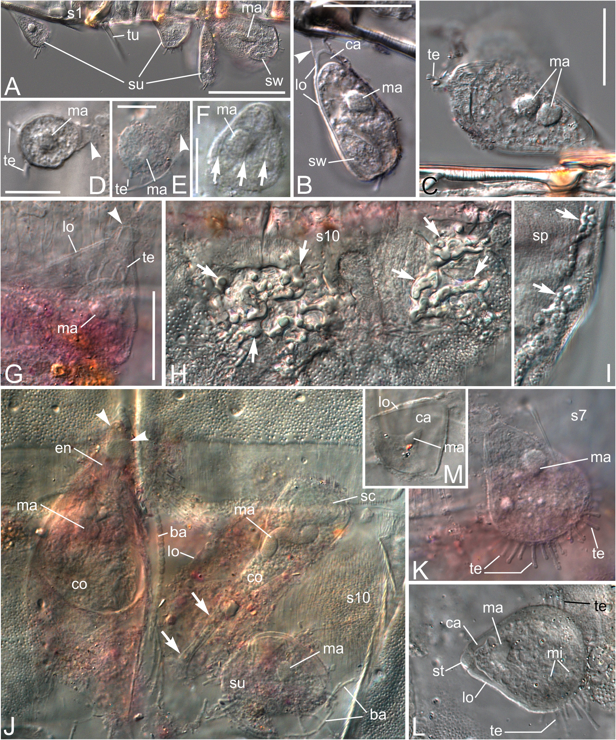
FIGURE 5. Setaphyes kielensis (A, ZMB 12489; B, ZMB 12500; C, ZMB 12499; D, ZMB 12487; J, ZMB 12486; L, ZMB 12397; M, ZMB 12369) and S. dentatus (E–G, K, ZMB 12503; F, H, I, ZMB 12494) with specimens of Trematosoma husselae sp. nov. (A–G, J–M), Cothurnia buetschlii (J), and a case of fungus or developmental artefact (H, I, ZMB 12494), DIC. A. Segments 1–4, lateral and dorso-ventral view on the calyciform, dorso-ventrally compressed lorica with the zooid, ventrolateral view on basibiont. B. Epibiont with swarmer, stalk, and lorica in optical section. C. Epibiont with two macronuclei not fusing at any focal level. D, E. Young, small, spherical epibionts with few tentacles, not all tentacles in focus. F. Young specimen on cuticle of basibiont with striation pattern on surface (arrows). G. Epibiont with tentacles in empty shell of suctorian epibiont. H, I. Segment 10, dorsally (H) and optical section (I), subcuticular fungus or internal sclerotisation artefact of the cuticle (arrows). J. Assemblage of epibionts on segment 10 of basibiont with C. buetschlii (contracted specimen left, extended specimen right), T. husselae sp. nov., and two kinds of filamentous bacteria. Arrows mark endostyle. K. Suctorian on segment 7 of S. dentatus. L, M. Specimens collected 1998, showing cellular substructures (L) and withdrawn zooid with minimum amount of cellular substructures (M). Extended focus images of six (D), eight (H, L), and three (K) images. Arrowheads mark stalks in B, D, E, G, and J. Abbreviations: ba, bacteria; ca, basal cavity in lorica; co, C. buetschlii; en, endostyle; lo, lorica; ma, macronucleus; s, segment, followed by segment number; sc, spherical cavity; sp, sperm; st, stalk; su, suctorian epibiont; sw, swarmer; te, tentacle; tu, tube on segment 2. Scale bar in A 50 µm, in B 20 µm, valid for B, G–J and M, in C 20 µm, valid for C, K, and L, and in D–F 10 µm.
Notes
Files
figure.png
Files
(13.8 MB)
| Name | Size | Download all |
|---|---|---|
|
md5:5621f7f530b3211dd9fc56f5e4122029
|
13.8 MB | Preview Download |