Published February 28, 2019
| Version v1
Figure
Open
Fig. 4 in Redescriptions of Euplotes encysticus and E. rariseta (Protist: Ciliophora: Euplotida)
Creators
- 1. Department of Environment and Energy Engineering, Kyungnam University, Changwon 51767, Republic of Korea & W&S Ecosystem Assessment Institute, Masanhappo-gu, Changwon 51745, Republic of Korea
- 2. Department of Environment and Energy Engineering, Kyungnam University, Changwon 51767, Republic of Korea
Description
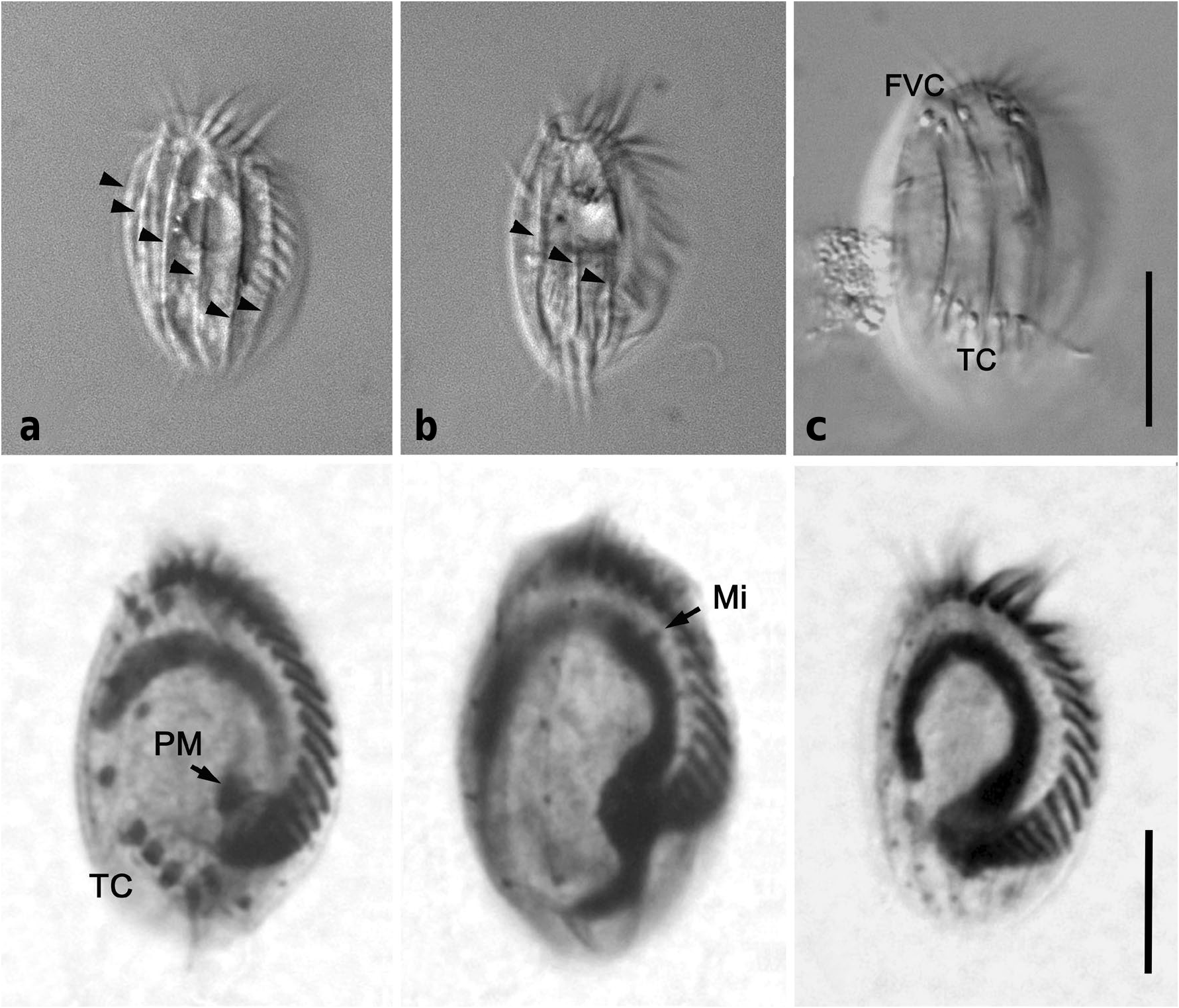
Fig. 4. Micrographs of Euplotes rariseta strain KM401 (a-c) in vivo and strain KM444 (d-f) after protargol impregnation. (a) Dorsal ridges (arrowheads) of E. rariseta, (b) ventral view showing longitudinal ridges (arrowheads), (c) ventral view showing FVC and TC, (d) ventral view showing AZM (adoral zone of membranelles), FVC (fronto-ventral cirri), TC (transvers cirri), PM (paroral membrane), (e) dorsal view showing dorsal kineties, (f) ventral view showing ventral kineties and Ma (macronucleus). Scale bars in (c) for (a-c) represent 20 μm and in (f) for (d-f) represent 10 μm.
Notes
Files
figure.png
Files
(2.4 MB)
| Name | Size | Download all |
|---|---|---|
|
md5:63f11ab144e31ef348b26a94f0bf4642
|
2.4 MB | Preview Download |