Published December 31, 2022
| Version v1
Figure
Open
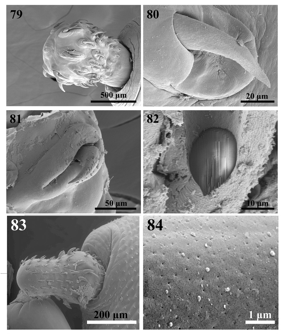
Figs 79–84 in Sem Study Of Hooks In The Acanthocephala With Emphasis On Structural-Functional Relationships
Creators
- 1. Institute of Parasitic Diseases, 11445 E. Via Linda 2-419, Scottsdale, Arizona 85259, USA
- 2. Department of Biology, Brigham Young University, 1114 MLBM, Provo, Utah 84602, USA. Deceased. Corresponding author
Description
Figs 79–84. SEM of proboscis and hooks of Pachysentis canicola (Oligacanthorhynchidae) (figs 79–82) and Corynosoma strumosum (Polymorphidae) (figs 83, 84): 79 — the proboscis of a female P. canicola showing hook arrangement and sensory pores at posterior proboscis and neck; 80 — an anterior hook deeply recessed in thick cuticular fold; 81 — a posterior hook also deeply recessed in a boat-like cuticular fold; 82 — a Gallium-cut cross section of a hook near its base showing the ventral protrusion as seen in other oligacanthorhynchid genera: Macracanthorhynchus and Nephridiacanthus; 83 — the proboscis of a specimen of C. strumosum showing its bare apical end and larger hooks at the bulge; 84 — a high magnification of a hook showing micropores.
Other
Published as part of Amin, O. M. & Heckmann, R. A., 2022, Sem Study Of Hooks In The Acanthocephala With Emphasis On Structural-Functional Relationships, pp. 265-284 in Zoodiversity 56 (4) on page 278, DOI: 10.15407/zoo2022.04.265, http://zenodo.org/record/7175553Files
figure.png
Files
(1.2 MB)
| Name | Size | Download all |
|---|---|---|
|
md5:a524dc3276f16ccf12793e854a74c03c
|
1.2 MB | Preview Download |
Linked records
Additional details
Related works
- Is part of
- Journal article: 10.15407/zoo2022.04.265 (DOI)
- Journal article: urn:lsid:plazi.org:pub:DD29FF86FFF3FFC7FF807328FFF48729 (LSID)
- Journal article: https://zenodo.org/record/7175553 (URL)