Test Dataset for Whole Slide Image Registration
Authors/Creators

- 1. BioImaging & Optics platform (BIOP), Faculty of Life Sciences (SV), Ecole Polytechnique Fédérale de Lausanne (EPFL), Lausanne, Switzerland
Description
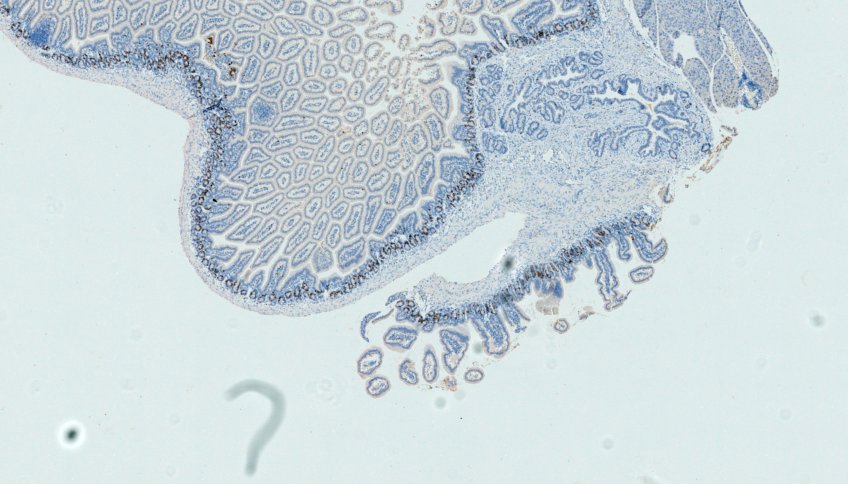
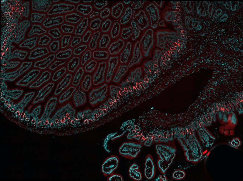
Mouse duodenum fixed in 4% PFA overnight at 4°C, processed for paraffin infiltration using a standard histology procedure and cut at 4 microns were dewaxed, rehydrated, permeabilized with 0.5% Triton X-100 in PBS 1x and stained with Azide - Alexa Fluor 555 (Thermo Fisher) to detect EdU and DAPI for nuclei. The images were taken using a Leica DM5500 microscope with a 40X N.A.1 objective (black&white camera: DFC350FXR2, pixel dimension: 0.161 microns). Next, the slide was unmounted and stained using the fully automated Ventana Discovery xT autostainer (Roche Diagnostics, Rotkreuz, Switzerland). All steps were performed on automate with Ventana solutions. Sections were pretreated with heat using the CC1 solution under mild conditions. The primary rat anti BrDU (clone: BU1/75 (ICR1), Serotec, diluted 1:300) was incubated 1 hour at 37°C. After incubation with a donkey anti rat biotin diluted 1:200 (Jackson ImmunoResearch Laboratories), chromogenic revelation was performed with DabMap kit. The section was counterstained with Harris hematoxylin (J.T. Baker) before a second round of imaging on DM5500 PL Fluotar 40X N.A.1.0 oil (color camera: DFC 320 R2, pixel dimension: 0.1725 microns). Before acquisition, a white-balance as well as a shading correction is performed according to Leica LAS software wizard. The fluorescence and DAB images were converted in ome.tiff multiresolution file with the kheops Fiji Plugin.
Sampled prepared in the EPFL histology core facility by Nathalie Müller and Gian-Filippo Mancini.
Associated documents:
- https://c4science.ch/w/bioimaging_and_optics_platform_biop/teaching/dab-intensity/
- https://imagej.net/plugins/bdv/warpy/warpy
This document contains a full QuPath project with an example of registered image.