Published October 31, 2006
| Version v1
Figure
Open
Figure 2 in A new species of the genus Lightiella: the first record of Cephalocarida (Crustacea) in Europe
Description
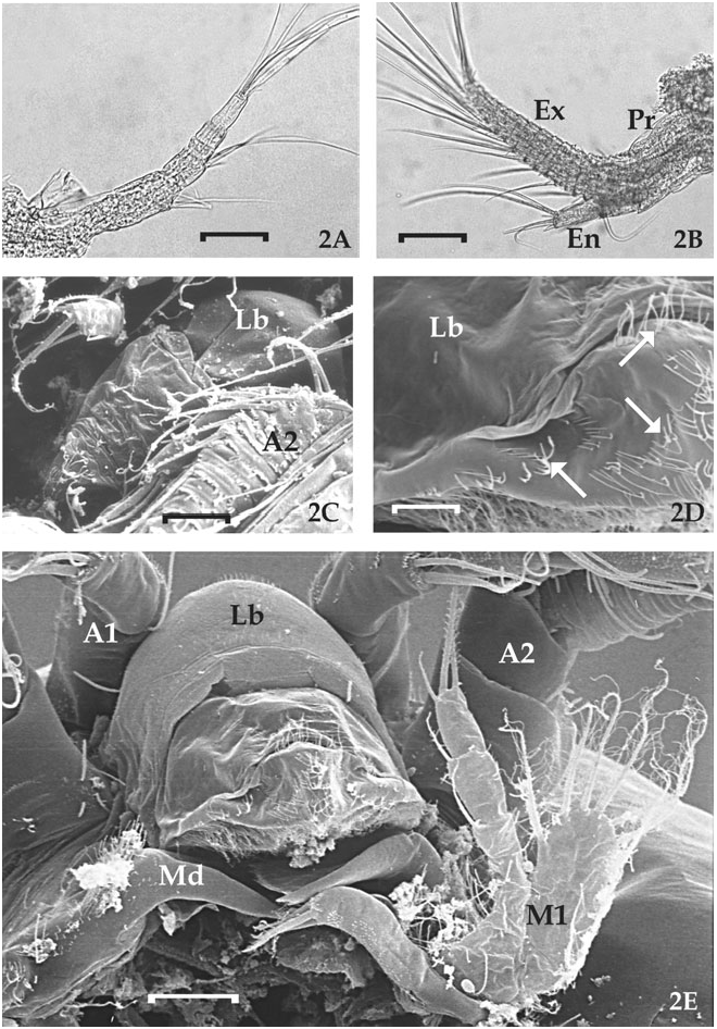
Figure 2. Light and scanning electron micrographs of first and second antennae and labrum of Lightiella magdalenina sp. nov. A, 6-segmented first antenna. Scale bar = 70 µm. B, second antenna with 2-segmented protopod (Pr), 2-segmented endopod (En) and 19-segmented exopod (Ex). Scale bar = 80 µm. C, ventral view of labrum (Lb), which appears rounded anteriorly and acutely triangular posteriorly; second antenna (A2). Scale bar = 45 µm. D, detail of the postero-ventral surface of labrum (Lb) covered by thin setae randomly distributed (arrows). Scale bar = 3 µm. E, posterior view of cephalon separated by the remaining part of the body at the level of the second maxilla. First antenna (A1), second antenna (A2), labrum (Lb), unsegmented mandible (Md), first maxilla (M1). Scale bar = 10 µm.
Notes
Files
figure.png
Files
(673.3 kB)
| Name | Size | Download all |
|---|---|---|
|
md5:e4d996fa76583263a103a160f2315b50
|
673.3 kB | Preview Download |
Linked records
Oops! Something went wrong while fetching results.