Published December 31, 2015
| Version v1
Figure
Open
FIGURE 2 in Description and phylogeny of a new prostomatid, Metacystis similis nov. spec. (Protista, Ciliophora) from the East China Sea
Description
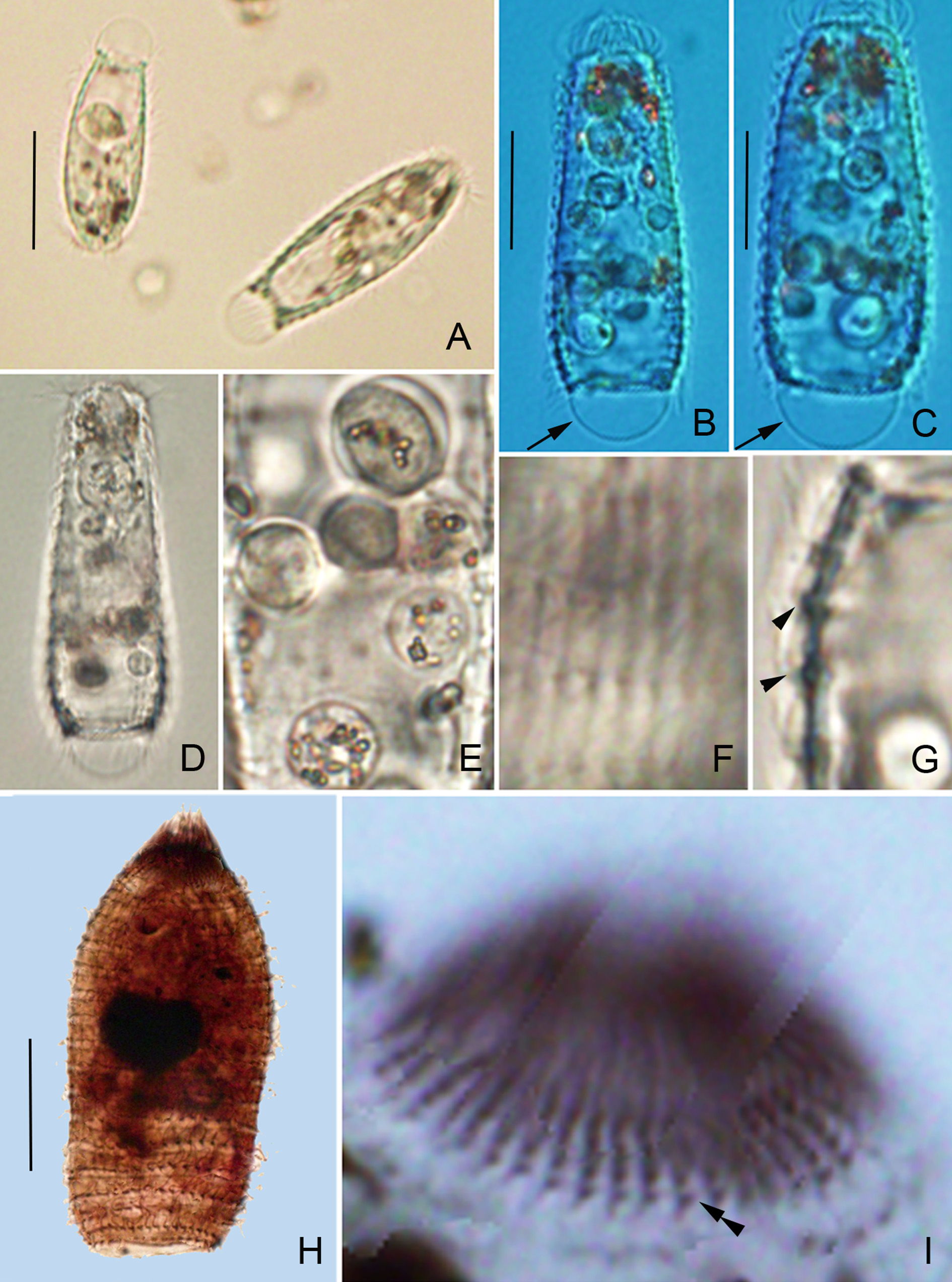
FIGURE 2. Photomicrographs of Metacystis similis nov. spec. from living specimens (A–G) and after protargol staining (H–I). A. Living individuals under low magnification. B-D. Lateral view of two typical individuals, arrows show the caudal vacuole. E. Food vacuoles and granules. F. Detail of the body surface; rectangular pellicle meshes. G. Detail of the pellicle structure; ridges on the cell surface (arrowheads). H. General infraciliature. I. Buccal area, double arrowhead refers to the circumoral ciliature. Scale bars: 50 µm (Fig. A); 30 µm (Figs. B, C, H).
Notes
Files
figure.png
Files
(9.6 MB)
| Name | Size | Download all |
|---|---|---|
|
md5:9df70d3f0a165a13c20d5ffa68476e26
|
9.6 MB | Preview Download |