Published December 31, 2009
| Version v1
Figure
Open
FIGURE 11 in Description of the first cryptobranch onchidoridid Onchimira cavifera gen. et sp. nov., and of three new species of the genera Adalaria Bergh, 1879 and Onchidoris Blainville, 1816 (Nudibranchia: Onchidorididae) from Kamchatka waters
Description
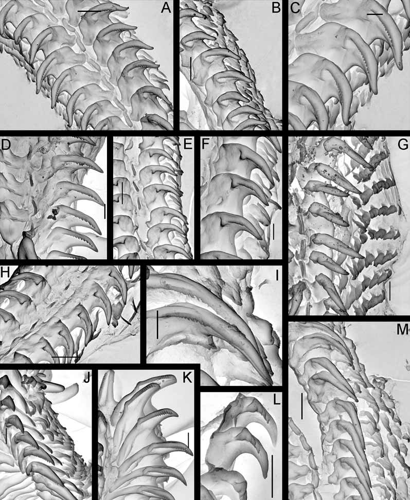
FIGURE 11. Radulae of species of the genus Adalaria, scanning electron micrographs. A–C, Adalaria slavi sp. nov., paratype ZMMU Lc-37457, living specimen, 18 mm length; A. Middle part of the radula; B. Several middle rows showing outer laterals; C. Enlarged first laterals showing cusp denticle pattern; D. Adalaria slavi sp. nov., paratype ZMMU Lc-37460, juvenile, 7 mm, middle part of the radula; E–F, Adalaria olgae sp. nov., paratype ZMMU Lc-37454, living specimen, 9 mm length; E. Middle part of the radula; F. Few enlarged middle rows showing cusp denticles pattern of the first laterals and outer lateral teeth; G. Adalaria proxima (Alder & Hancock, 1854), ZMMU, not registered, Barents Sea, Dalne-Zelenetskaya Bay, living non-mature specimen with poorly differentiated reproductive system and smooth first lateral teeth, 15 mm length, intertidal, middle part of the radula; H. Adalaria olgae sp. nov., paratype, ZMMU Lc- 37455, living specimen, 10 mm length, middle part of the radula; I. Adalaria tschuktschica Krause, 1885, ZMMU, not registered, preserved specimen, 8 mm length, Chukchi Sea, Vrangel. Id., from 7 m depth, close up of the first laterals from the middle part of the radula showing pattern of cusp denticles; J–K, Adalaria jannae Millen 1987, ZMMU, not registered, living specimen, 8 mm length, Starichkov Island; J. Middle part of the radula, showing outer lateral teeth; K. Close up of the first laterals showing pattern of cusp denticles; L. Adalaria proxima (Alder et Hancock, 1854), ZMMU, not registered, juvenile specimen, 5 mm length, White Sea; M. Adalaria tschuktschica, middle part of the radula. Scale bars: A―50 μm, B―50 μm, C―20 μm, D―20 μm, E―50 μm, F―20 μm, G―30 μm, H―50 μm, I―20 μm, J―20 μm, K―20 μm, L―30 μm, M―50 μm. Photos: Alexander Martynov.
Notes
Files
figure.png
Files
(767.2 kB)
| Name | Size | Download all |
|---|---|---|
|
md5:b9e02748337a37aabffd24fd87870bd6
|
767.2 kB | Preview Download |