Published September 11, 2025
| Version v2
Image
Open
NFDI4BIOIMAGE Calendar March 2025

Description
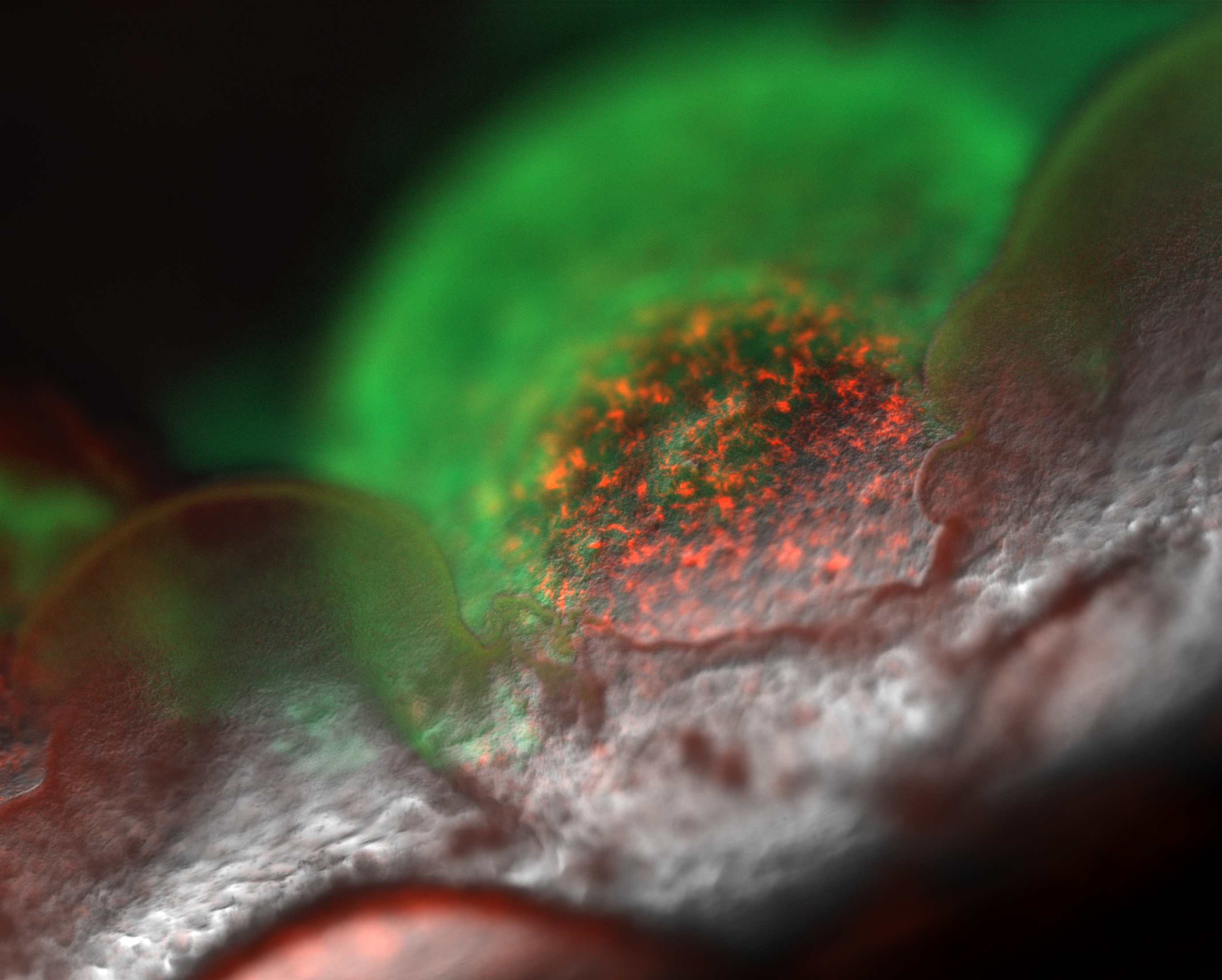
Raw microscopy image from the NFDI4Bioimage calendar March 2025.
The image shows 125x magnified microscopic details of a biofilm formed by Pseudomonas fluorescence on the surface of a liquid culture medium. The culture was inoculated with a cellulose-overexpressing and surface-colonizing mScarlet-tagged wild type and a GFP-tagged mutant that is unable to colonize the surface. The biofilm can collapse over time due to its own mass, so that new strategies have to be developed and thus a life cycle emerges.
Image Metadata (using REMBI template):
| Study | |
| Study description | Biofilm formation |
| Study Component | |
| Imaging method | Stereo microscopy |
| Biosample | |
| Biological entity | Bacteria |
| Organism | Pseudomonas fluorescence |
| Specimen | |
| Signal/contrast mechanism | Relief, fluorescence |
| Channel 1 - content | Relief, grey |
| Channel 1 - biological entity | Details of the biofilm in transmitted light |
| Channel 2 - content | mScarlet, red |
| Channel 2 - biological entity | WT over-expressing cellulose and colonizing the surface |
| Channel 3 - content | GFP, green |
| Channel 3 - biological entity | ∆wss mutant unable to colonize the surface |
| Image Acquisition | |
| Microscope model | Zeiss Axio Zoom V16 |
| Image Data | |
| Magnification | 125x |
| Objective | PlanNeoFluar Z 1.0x |
| Dimension extents | x: 2752, y: 2208 |
| Pixel size description | 0.91 µm x 0.91 µm |
| Image area | 2500µm x 2500µm |
| Submitted via NFDI4BIOIMAGE |
Files
image_march.png
Files
(46.3 MB)
| Name | Size | Download all |
|---|---|---|
|
md5:a0806f3d96cb9616a596ebf1844a709d
|
9.3 MB | Preview Download |
|
md5:c072912dcf375377a3d0ad033463947e
|
37.1 MB | Download |
Additional details
Related works
- Is identical to
- Image: 10.57860/min_img_000010 (DOI)