NFDI4BIOIMAGE Calendar December 2025

- 1. Institute of Microbiology, Heinrich Heine University Düsseldorf, Germany
Contributors
Data curator:
-
1.
 Heinrich Heine University Düsseldorf
-
2.
NFDI4BIOIMAGE
Description
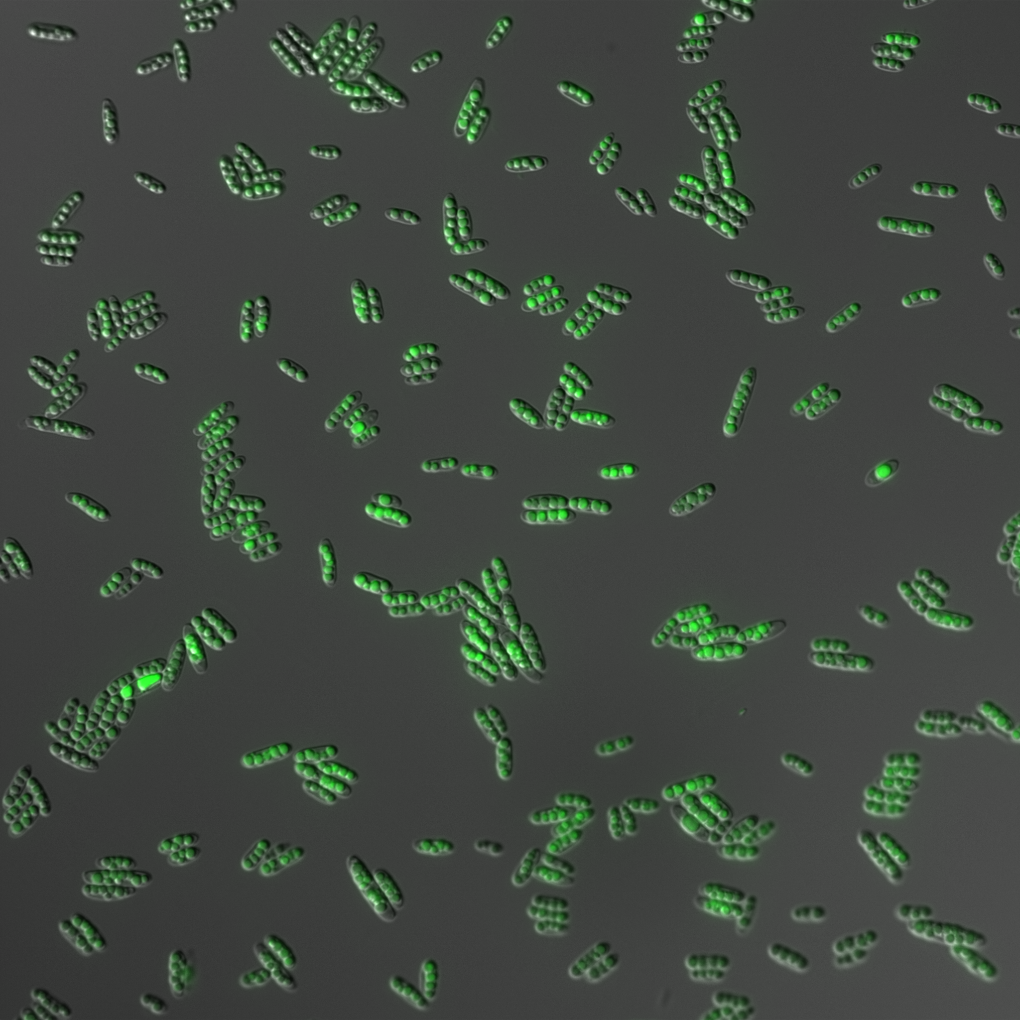
Image from the NFDI4BIOIMAGE Calendar December 2025.
The microscopic image shows yeast cells of the fungal model Ustilago maydis that produce single cell oil at nitrogen-starvation conditions. The genetically engineered cells are packed with oil droplets that were visualized by BODIPY staining. The study was conducted in the framework of the BioSC project "NextVegOil".
Image Metadata (using REMBI template):
|
Study |
|
|
Study type |
Visualisation of microbial oil in the fungus Ustilago maydis |
|
Study Component |
|
|
Imaging method |
Wide field whole organism microscopy |
|
Biosample |
|
|
Biological entity |
Ustilago maydis |
|
Organism |
Yeast cells (sporidia) |
|
Identity |
Ustilago maydis MB215 cyp1Δemt1Δ (published in https://doi.org/10.1128/AEM.71.6.3033-3040.2005) |
|
Intrinsic variable |
Glycolipid production has been ablated by genetic engineering |
|
Extrinsic variable |
BODIPY (4,4-Difluoro-1,3,5,7,8-Pentamethyl-4-Bora-3a,4a-Diaza-s-Indacene 493/503) staining |
|
Experimental variables |
Cultivation time |
|
Specimen |
|
|
Location within biosample |
Overview image with yeast cells from liquid culture at nitrogen-starvation condition |
|
Preparation method |
Living cells attached to agarose mounts |
|
Signal/contrast mechanism |
Differential interference contrast and fluorescence |
|
Channel 1 - content |
DIC |
|
Channel 1 - biological entity |
Intact yeast cells |
|
Channel 2 - content |
BODIPY 493/503 |
|
Channel 2 - biological entity |
Intracellular lipid droplets |
|
Image acquisition |
|
|
Instrument attributes |
Zeiss Axio Observer.Z1; Prime BSI express; solid-state laser 488 nm |
|
Submitted via NFDI4BIOIMAGE |
|
Files
image_december.png
Files
(5.0 MB)
| Name | Size | Download all |
|---|---|---|
|
md5:a0073ead8be757dcbc12b16122588e87
|
5.0 MB | Preview Download |