NFDI4BIOIMAGE Calendar September 2025
Authors/Creators

- 1. Leibniz Institute for Neurobiology, Magdeburg, Germany
- 2. Combinatorial NeuroImaging Core Facility, Leibniz Institute for Neurobiology, Magdeburg, Germany
- 3. Institute for Molecular and Clinical Immunology and Service Unit Multiparametric Bioimaging and Cytometry, University of Magdeburg, Germany
- 4. Institute of Materials, Technologies and Mechanics, University of Magdeburg, Germany
Contributors
Data curator:
-
1.
 Heinrich Heine University Düsseldorf
-
2.
NFDI4BIOIMAGE
Description
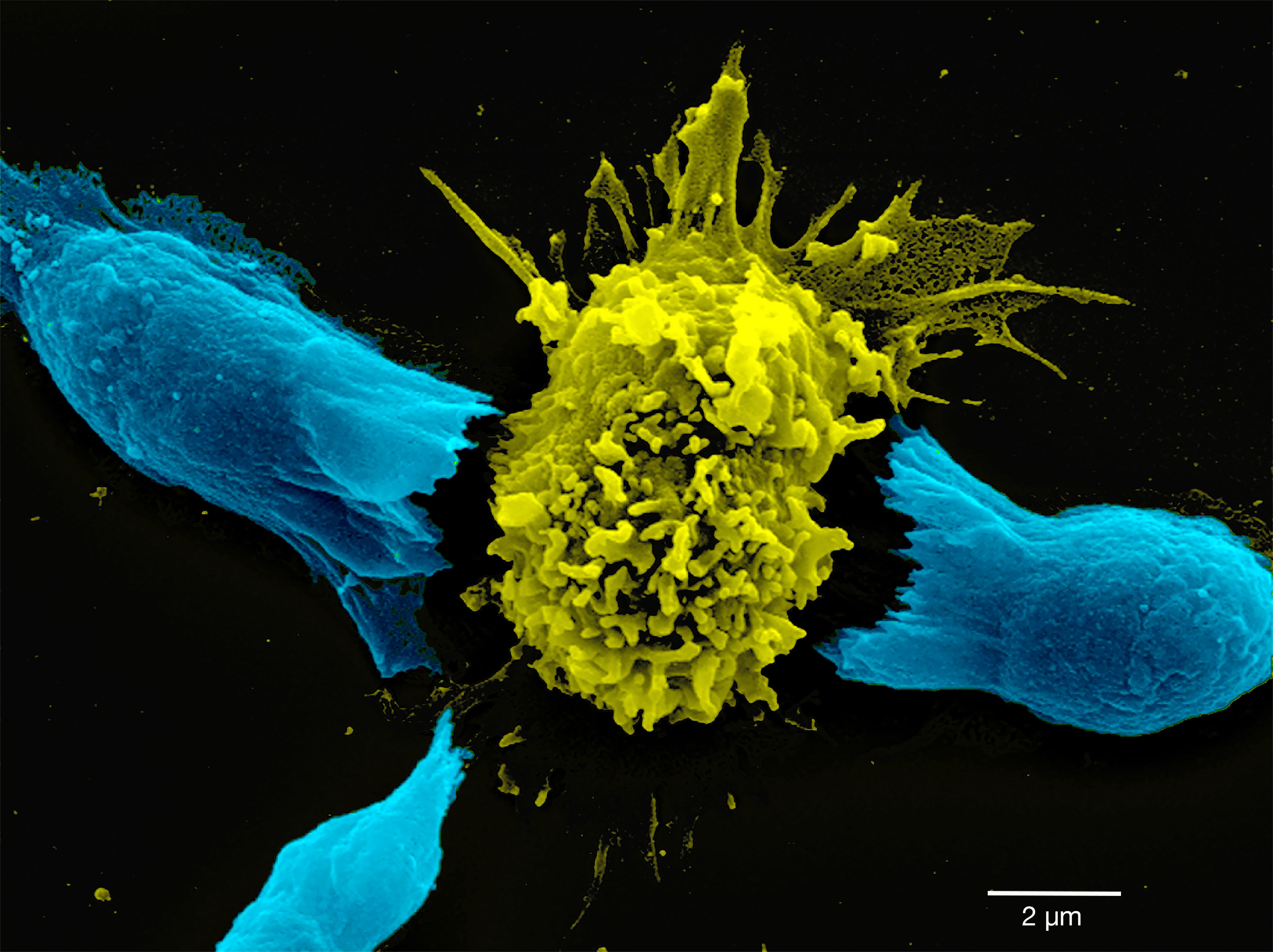
Image from the NFDI4BIOIMAGE Calendar September 2025.
The scanning electron micrograph shows the approach of T-lymphocytes (Jurkat cells; cyan) to an antigen-presenting B cell (Raji cell; yellow) in the center. The image was taken as part of the research work of the CRC 854, which focused on molecular processes that regulate inter- and intracellular communication within the immune system.
Image Metadata (using REMBI template):
|
Study |
|
|
Study description |
Ultrastructure of the immune synapse |
|
Study type |
Research project within DFG CRC 854 (Molecular organisation of cellular communication within the immune system) |
|
Study Component |
|
|
Imaging method |
Scanning Electron Microscopy |
|
Biosample |
|
|
Biological entity |
Jurkat cell line E6.1 and Raji B cell lymphoma cell line |
|
Organism |
Homo sapiens |
|
Identity |
Z21_A1 |
|
Specimen |
|
|
Preparation method |
Cell lines were maintained in RPMI 1640 medium supplemented with 10% fetal calf serum (FCS; PAN Biotech), stable L-glutamine, penicillin (50 U/ml), and streptomycin (50 mg/ml) (Biochrom) in humidified 5% CO2 at 37°C. E6.1 cells were mixed at a 1:1 ratio with Raji B cells that had been pulsed with SEE (bacterial SAG staphylococcal enterotoxin E). After 10 min cells were plated on poly-L-lysine–covered slides at room temperature for 5 min and fixed for 10 min in PBS (pH 7.4) containing 1.5% PFA and 0.025% glutaraldehyde. Cryo-drying by critical point dryer (Leica EM CPD300) followed by sputtering with gold. |
|
Signal/contrast mechanism |
Detected secondary electrons |
|
Channel 1 - content |
Jurkat cell line E6.1 (artificial color table, cyan) |
|
Channel 1 - biological entity |
Surface of Jurkat cells |
|
Channel 2 - content |
Raji B cell lymphoma cell line (artificial color table, yellow) |
|
Channel 2 - biological entity |
Surface of a Raji B cell |
|
Image acquisition |
|
|
Instrument attributes |
FEI XL30 FEG ESEM |
|
Image acquisition parameters |
10 keV, Magnification 6500 x, Scale bar: 2 µm |
|
Submitted via NFDI4BIOIMAGE |
|
Files
Files
(5.1 MB)
| Name | Size | Download all |
|---|---|---|
|
md5:e01ad0296a5fbf910d1ce2c976c02267
|
5.1 MB | Download |