cytoplasm_nucleus_translocation_cells_8bip
Creators

Contributors
Data collector:
Description
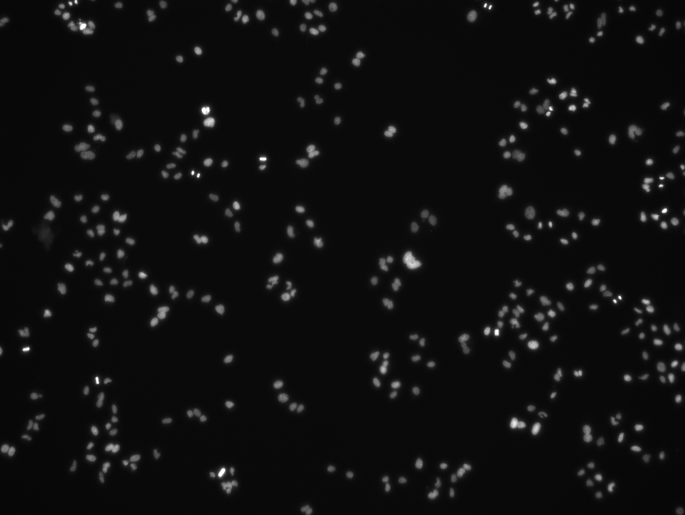
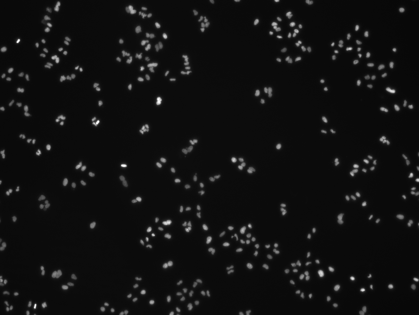
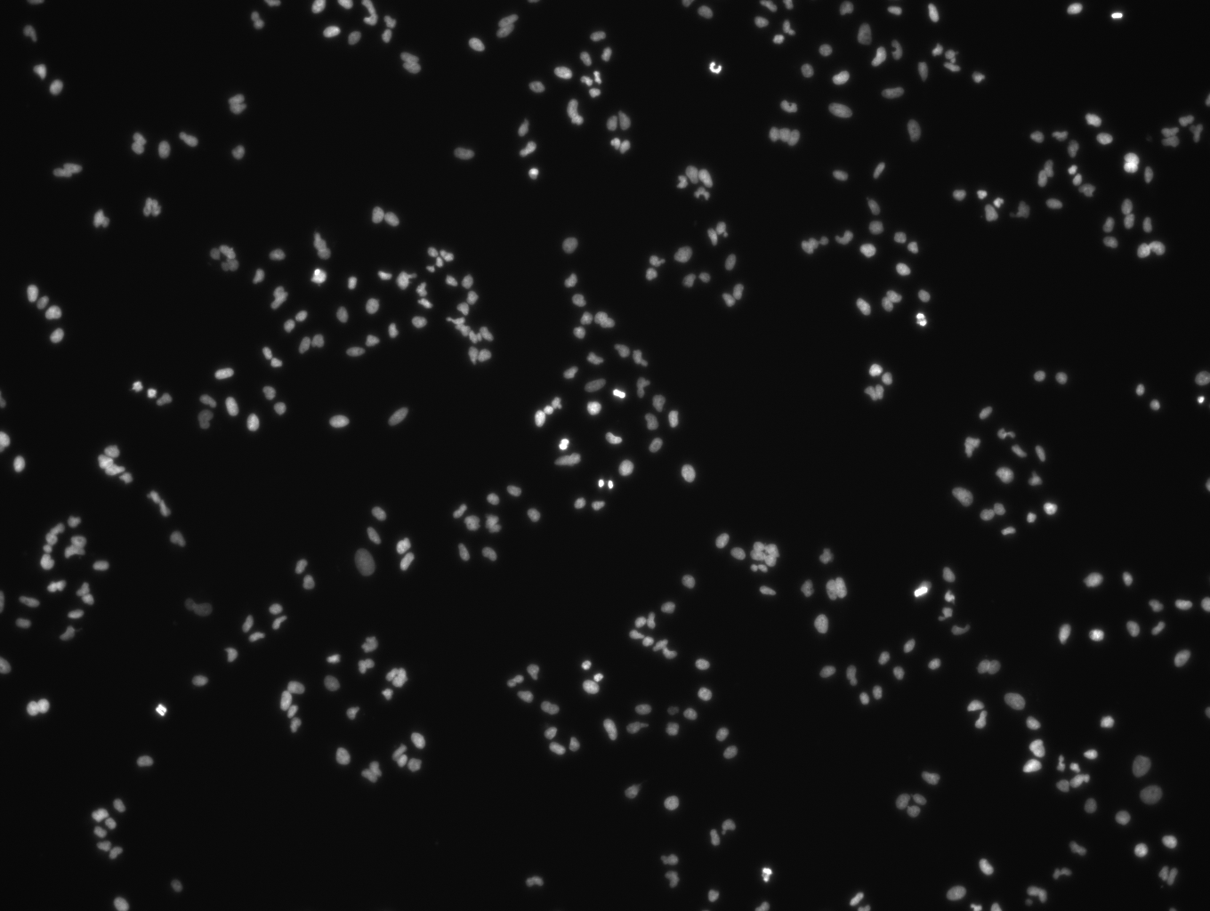
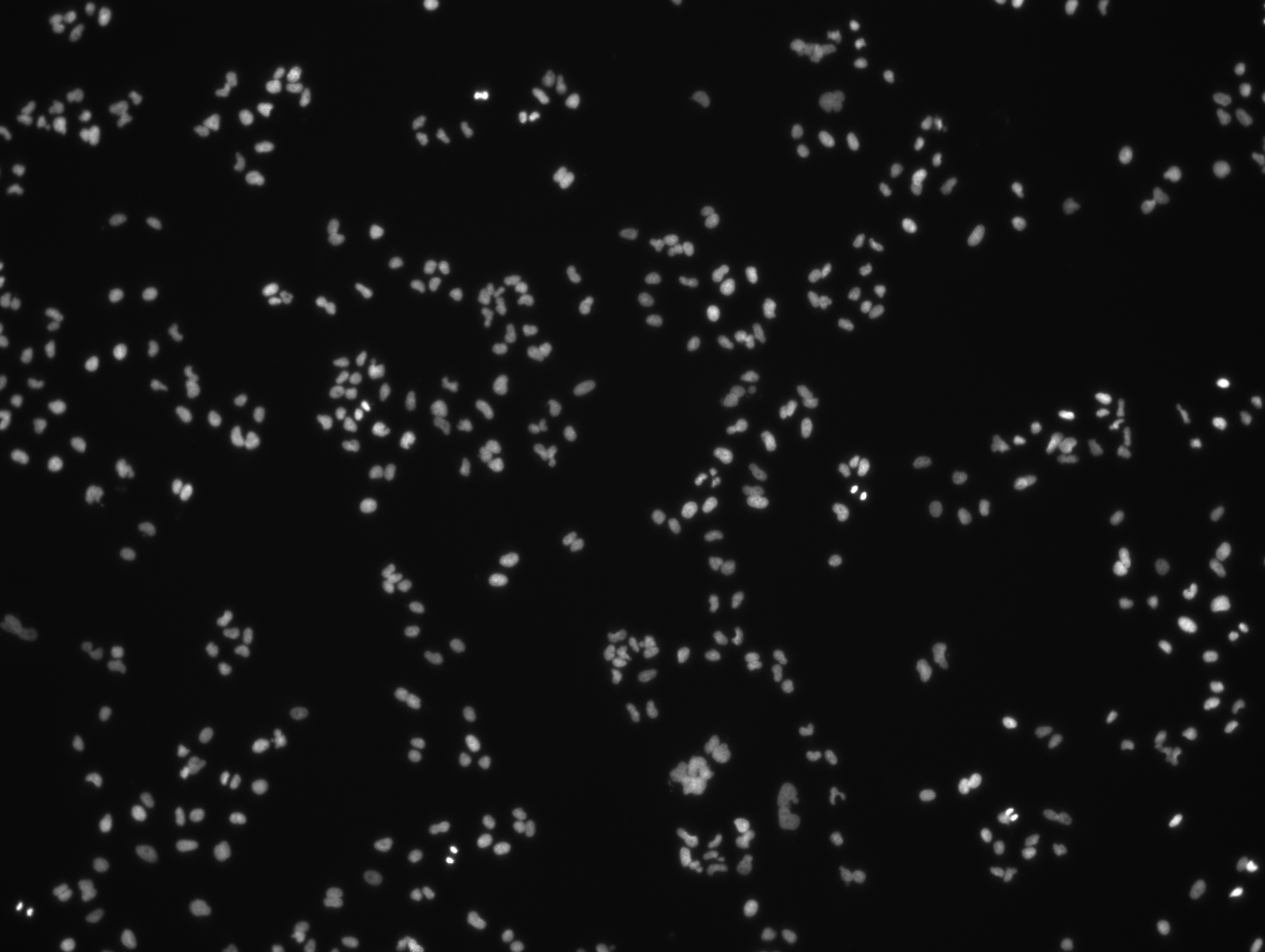
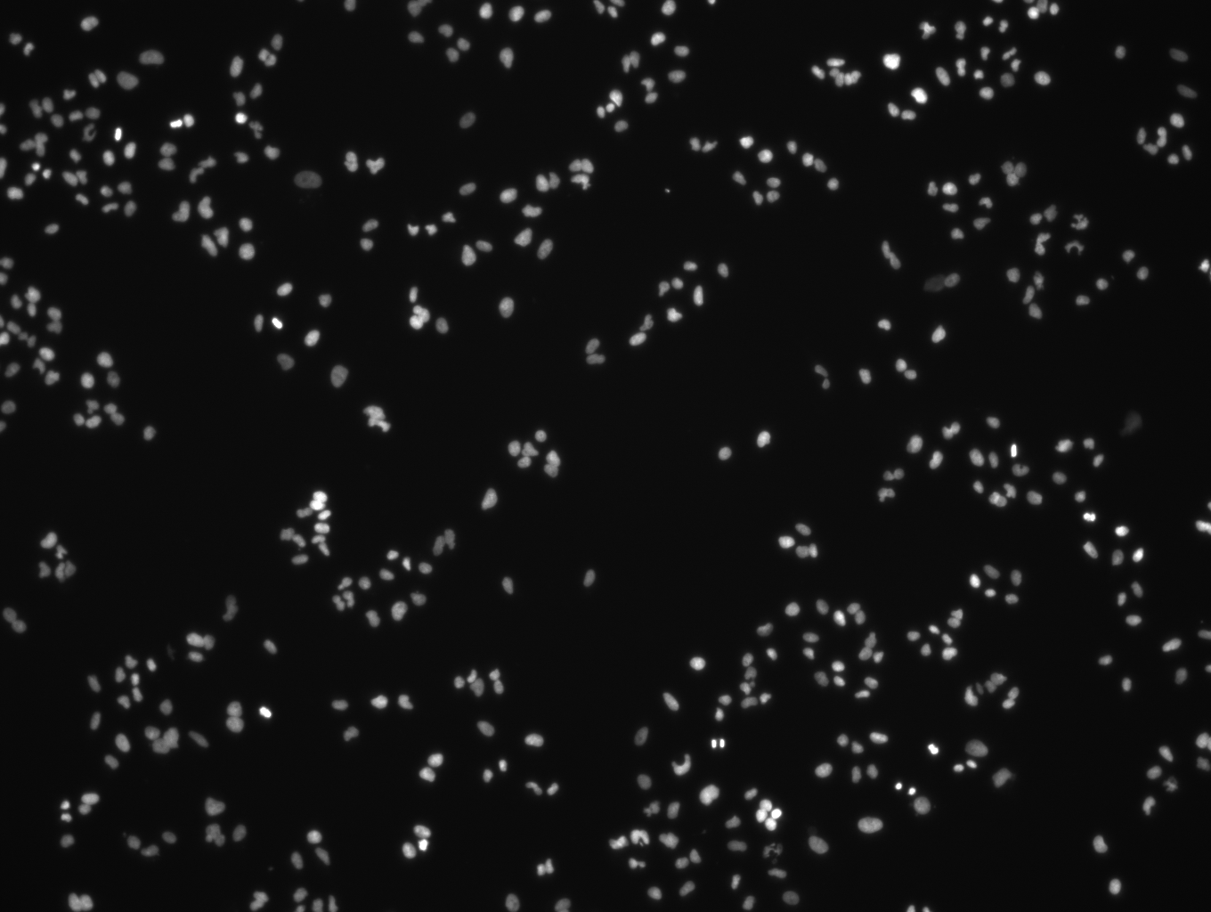
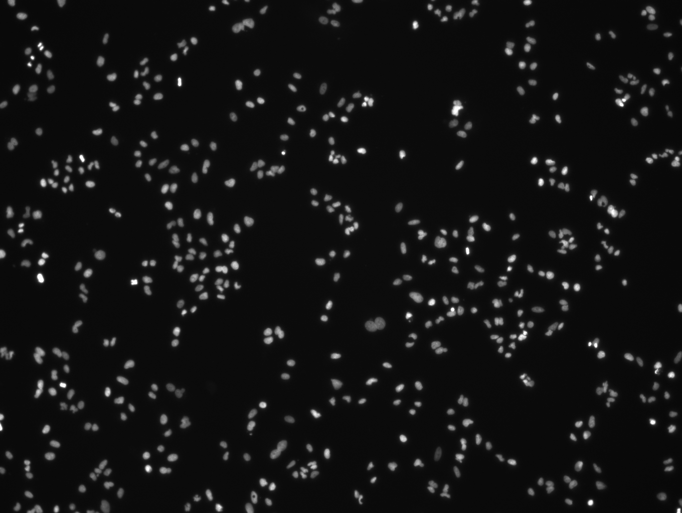
Sub-sample images: Images of cytoplasm to nucleus translocation of the transcription factor NFκB in MCF7 (human breast adenocarcinoma cell line) and A549 (human alveolar basal epithelial) cells in response to TNFα concentration. Images are at 10x objective magnification. The plate was acquired at Vitra Bioscience on the CellCard reader. For each well there is one field with two images: a nuclear counterstain (DAPI) image and a signal stain (FITC) image. Image size is 1360 x 1024 pixels. Images are in 8-bit BMP format.
Source: https://bbbc.broadinstitute.org/BBBC014
Citation: Ljosa, Vebjorn, Katherine L. Sokolnicki, and Anne E. Carpenter. "Annotated high-throughput microscopy image sets for validation." Nature methods 9.7 (2012): 637-637. https://doi.org/10.1038/nmeth.2083
Notes
Files
Files
(12.5 MB)
| Name | Size | Download all |
|---|---|---|
|
md5:ee0aff2c62cd6cb917759899e1882235
|
1.4 MB | Download |
|
md5:f4142446266c74148c955c79f58a8666
|
1.4 MB | Download |
|
md5:2f8753833a5f4a4353168590043100c2
|
1.4 MB | Download |
|
md5:33f2a36186da90a576b7601b2c1fc7dd
|
1.4 MB | Download |
|
md5:3b75fcfcc3dcc0a914cf9a0d0614e5c8
|
1.4 MB | Download |
|
md5:f729afb8a289696207ee7a35dd9814ce
|
1.4 MB | Download |
|
md5:2855cfc34cbbcb075c10753ba4931edd
|
1.4 MB | Download |
|
md5:2b6195275c6d18b5ffef09113aa86d41
|
1.4 MB | Download |
|
md5:870676c7716d1c977485d351f1f09f2c
|
1.4 MB | Download |