Published June 29, 2006
| Version v1
Figure
Open
FIGURE 1 in Pseudourostyla pelotensis sp. nov. (Ciliophora, Stichotrichia, Urostylida): a new psammophilic ciliate from the southern Brazil
Description
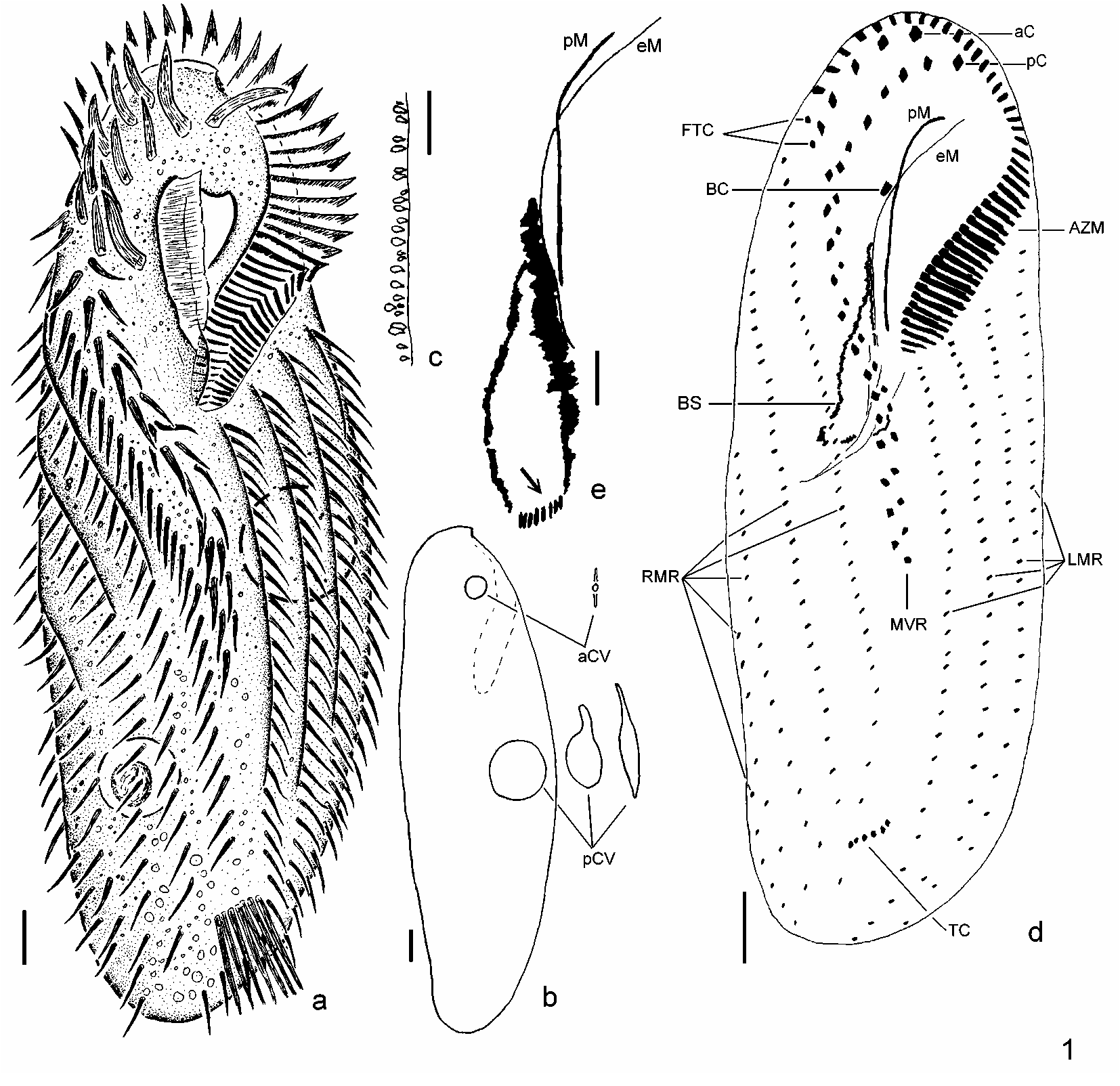
FIGURE 1. Schematic drawings of Pseudourostyla pelotensis sp. nov. a. Living specimen in ventral view; b. Scheme of living specimen showing the contractile vacuoles and their aspect during pulsation; c. Extrusomes aligned close to the pelicle, as seen in specimens from protargol slides; d. Ventral region of protargolimpregnated specimen; e. Bowshaped structure associated with the undulating membranes. Arrow marks new adoral membranelles in formation. aC — anterior corona; aCV — anterior contractile vacuole; AZM — adoral zone of membranelles; BC — buccal cirrus; BS — bowlike structure; eM — endoral membrane; FTC — frontotermial cirri; LMR — left marginal cirral rows; MVR — midventral cirral row; pC — posterior corona; pCV — posterior contractile vacuole; pM — paroral membrane; RMR — right marginal cirral rows; TC — transverse cirri. Scale bars: 10 µm.
Other
Published as part of Paiva, Thiago Da Silva & Silva-Neto, Inácio Domingos Da, 2006, Pseudourostyla pelotensis sp. nov. (Ciliophora, Stichotrichia, Urostylida): a new psammophilic ciliate from the southern Brazil, pp. 43-58 in Zootaxa 1247 (1) on page 46, DOI: 10.11646/zootaxa.1247.1.4, http://zenodo.org/record/10087469Files
figure.png
Files
(199.4 kB)
| Name | Size | Download all |
|---|---|---|
|
md5:92897ce62c4bea61f40c7508d010e56a
|
199.4 kB | Preview Download |
Linked records
Additional details
Related works
- Is cited by
- Taxonomic treatment: http://treatment.plazi.org/id/03C487CF6F72FFD60F0769213AA5F804 (URL)
- Is part of
- Journal article: 10.11646/zootaxa.1247.1.4 (DOI)
- Journal article: urn:lsid:plazi.org:pub:FFFDFFB76F70FFDF0E0F683A3E2AFF6B (LSID)
- Journal article: http://publication.plazi.org/id/FFFDFFB76F70FFDF0E0F683A3E2AFF6B (URL)
- Journal article: https://zenodo.org/record/10087469 (URL)