Published March 16, 2023
| Version v1
Figure
Open
FIGURE 6 in Three interesting fungi from American bullfrog larvae (Rana catesbeiana) in Yunnan, China
Creators

- 1. Center for Yunnan Plateau Biological Resources Protection and Utilization, College of Biological Resource and Food Engineering, Qujing Normal University, Qujing, Yunnan 655011, People's Republic of China & Master of Science Program in Applied Microbiology (International Program), Faculty of Science, Chiang Mai University, Chiang Mai 50200, Thailand & erfu20170431@gmail.com; https://orcid.org/0000-0003-2385-6402
- 2. Center for Yunnan Plateau Biological Resources Protection and Utilization, College of Biological Resource and Food Engineering, Qujing Normal University, Qujing, Yunnan 655011, People's Republic of China & reuven0319@gmail.com; https://orcid.org/0000-0001-7283-7596
- 3. Center for Yunnan Plateau Biological Resources Protection and Utilization, College of Biological Resource and Food Engineering, Qujing Normal University, Qujing, Yunnan 655011, People's Republic of China & saowaluckfai@gmail.com; https://orcid.org/0000-0002-4706-6547
- 4. Center for Yunnan Plateau Biological Resources Protection and Utilization, College of Biological Resource and Food Engineering, Qujing Normal University, Qujing, Yunnan 655011, People's Republic of China & cicidaidongqin@gmail.com; https://orcid.org/0000-0001-8935-8807
- 5. Center of Excellence in Fungal Research, Mae Fah Luang University, Chiang Rai 57100, Thailand & gaoying@mail.kib.ac.cn; https://orcid.org/0000-0001-8671-1978
- 6. Department of Biology, Faculty of Science, Chiang Mai University, Chiang Mai, Thailand & itthayakorn.p@cmu.ac.th; https://orcid.org/0000-0003-3376-4376
- 7. Center for Yunnan Plateau Biological Resources Protection and Utilization, College of Biological Resource and Food Engineering, Qujing Normal University, Qujing, Yunnan 655011, People's Republic of China & National Institute of Fundamental Studies (NIFS), Sri Lanka & samanthakarunarathna@gmail.com; https://orcid.org/0000-0001-7080-0781
Description
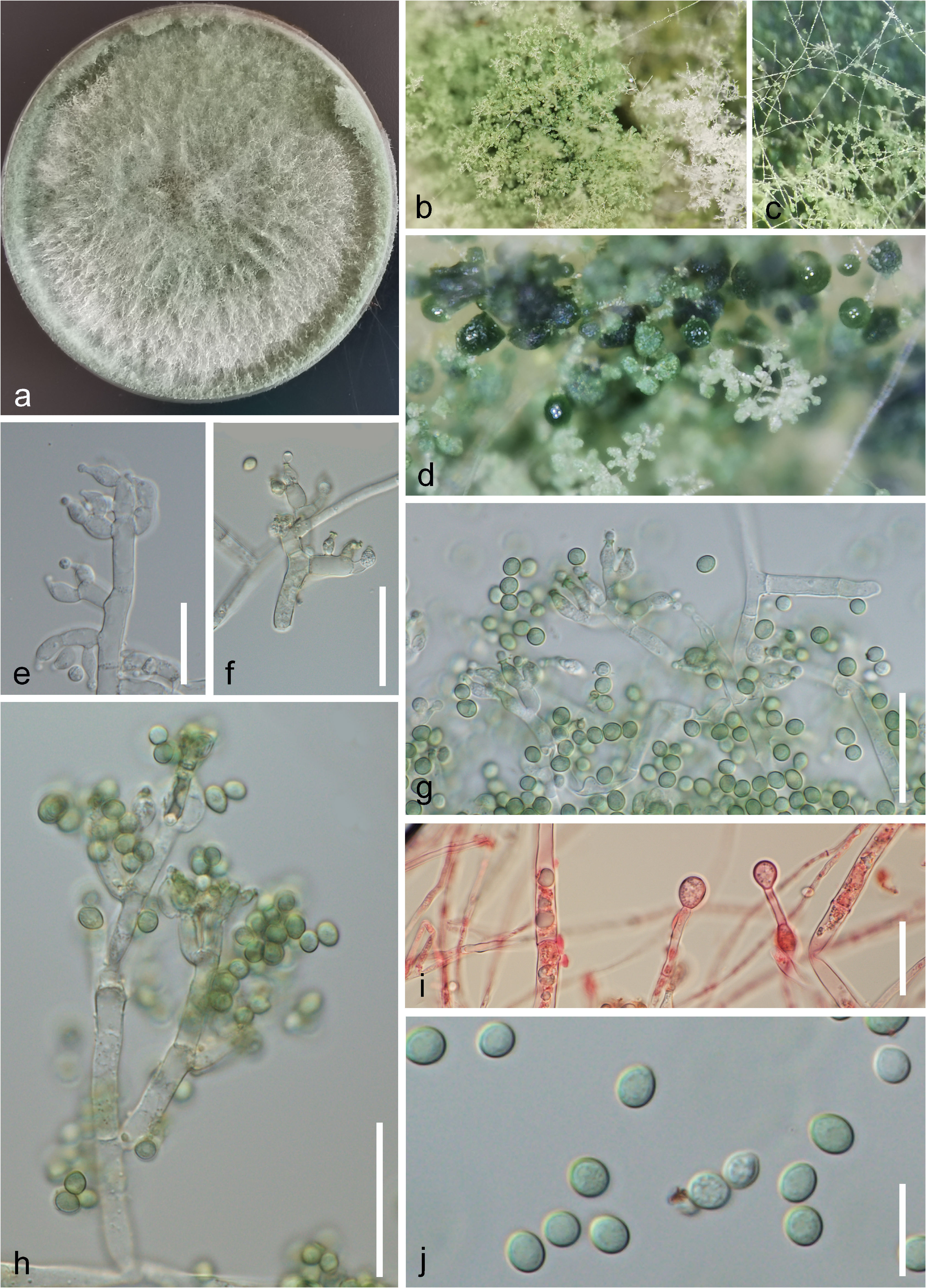
FIGURE 6 Trichoderma virens (HKAS 125765). a Colony on PDA for one month. b–d Close-up of colonies. e, f Undeveloped conidia and conidiogenous cells. g, h Mature conidia with conidiogenous cells. i Chlamydospores stained by congo red agents. j Conidia. Scale bars: g, h = μm; i = 20 μm; e, f = 15 μm; j = 10 μm.
Notes
Files
figure.png
Files
(29.1 MB)
| Name | Size | Download all |
|---|---|---|
|
md5:4e8c8da8ed6e3c028a5df006712be43b
|
29.1 MB | Preview Download |