Published October 1, 2018
| Version v1
Figure
Open
Fig. 5 in The Plasticity And Morphofunctional Organization Of The Digestive System Of Waders (Charadrii) As Migrants
Creators
Description
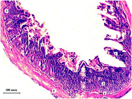
Fig. 5. The wall of the cecum Philomachus pugnax, the area of the body, cross cut. Histopreparation (hematoxylin and eosin, х100). 1 — mucosal plates; 2 — crypt; 3 — lymphoid tissue; 4 — submucosal basis; 5 — muscle.
Notes
Files
figure.png
Files
(363.7 kB)
| Name | Size | Download all |
|---|---|---|
|
md5:a82d3dafa7b9df8b7c8aa5c6033f1b4d
|
363.7 kB | Preview Download |
Linked records
Additional details
Related works
- Is part of
- Journal article: 10.2478/vzoo-2018-0043 (DOI)
- Journal article: urn:lsid:plazi.org:pub:992FFF91FF9AFFCAFF87FFB78724CC1D (LSID)
- Journal article: https://zenodo.org/record/6454887 (URL)