Published October 30, 2009
| Version v1
Figure
Open
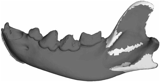
Figure 2 in Mandibular biomechanics of Crocuta crocuta, Canis lupus, and the late Miocene Dinocrocuta gigantea (Carnivora, Mammalia)
Creators
Description
Figure 2. Muscle attachment sites on the mandible finite element models, with Crocuta crocuta as an example. The light areas on top of the ascending ramus and in the mandibular fossa are attachment sites for the temporalis. The light area on the angular process is the attachment site of the masseter. The internal pterygoid attachment (not shown) is on the medial side of the angular process.
Notes
Files
figure.png
Files
(68.0 kB)
| Name | Size | Download all |
|---|---|---|
|
md5:eb100b7be40420e33fa7a40b2a8de2a7
|
68.0 kB | Preview Download |
Linked records
Additional details
Related works
- Is part of
- Journal article: 10.1111/j.1096-3642.2009.00555.x (DOI)
- Journal article: urn:lsid:plazi.org:pub:3B41FFF0FFA5FFF17E05FFDAC146FFE4 (LSID)
- Journal article: https://zenodo.org/record/5438190 (URL)