Published October 11, 2013
| Version v1
Figure
Open
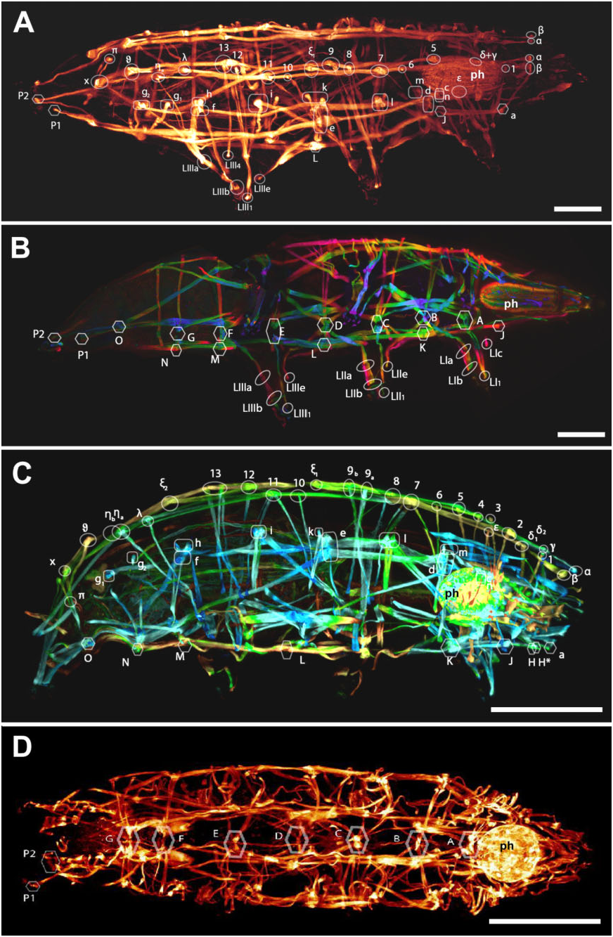
Figure 3. A, B in Somatic musculature of Tardigrada: phylogenetic signal and metameric patterns
Description
Figure 3. A, B, musculature of Milnesium cf. tardigradum (A, dorso-lateral view; B, ventro-lateral view). C, D, musculature of Acutuncus antarcticus (C, lateral view; D, ventro-lateral view). Ph, pharynx. Letters and numbers identify the muscle attachment points (see text). Nodes of ventral muscle groups are marked by hexagons; attachment points and nodes of lateral muscle group are marked by squares; attachment points and nodes of dorsal muscle group are marked by circles. A–D, CSLM, maximum projection. B, C, colour coded by depth. Scale bars: A–D = 50 μm.
Notes
Files
figure.png
Files
(1.7 MB)
| Name | Size | Download all |
|---|---|---|
|
md5:8adc3d7b0d055d63ad06fbe282d8a2cf
|
1.7 MB | Preview Download |
Linked records
Additional details
Related works
- Is part of
- Journal article: 10.1111/zoj.12079 (DOI)
- Journal article: urn:lsid:plazi.org:pub:FFDAFFD19A3EFFA1FFDAAE28A15CFFCB (LSID)
- Journal article: https://zenodo.org/record/5291175 (URL)