Figure 16 in New contributions to the marine benthic ciliates from the Antarctic area, including description of seven new species (Protozoa, Ciliophora)
Creators
Description
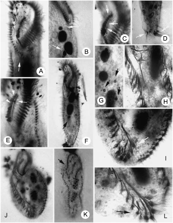
Figure 16. Photomicrographs of (A–F) Amphisiella antarctica nov. spec. and (G–L) Hemigastrostyla szaboi nov. spec. after protargol impregnation. (A) Ventral view of buccal region, to show the ventral row (arrow); (B) macro- and micronuclei (arrows); (C, E) buccal area, to show the argentophilic granules along the undulating membranes (arrows and arrowheads); (D) caudal portion, to show the transverse and caudal cirri; (F) ventral view, arrow marks the micronucleus; (G) nuclear apparatus, arrows indicate the micronuclei; (H) to show the fibres associated with the marginal cirri; (I) ventral view of posterior cell end, to show the transverse cirri, note that there is no gap between right marginal row and the transverse cirri; (J) ventral view, to show the general appearance of infraciliature; (K) ventral view, to show a slender form, arrow indicates the distal end of adoral zone of membranelles; (L) ventral view of caudal region, arrow marks the transverse cirri.
Notes
Files
figure.png
Files
(483.0 kB)
| Name | Size | Download all |
|---|---|---|
|
md5:cfb62c15cd9ea291d5e5176b67d17024
|
483.0 kB | Preview Download |