Figure 8 in New contributions to the marine benthic ciliates from the Antarctic area, including description of seven new species (Protozoa, Ciliophora)
Creators
Description
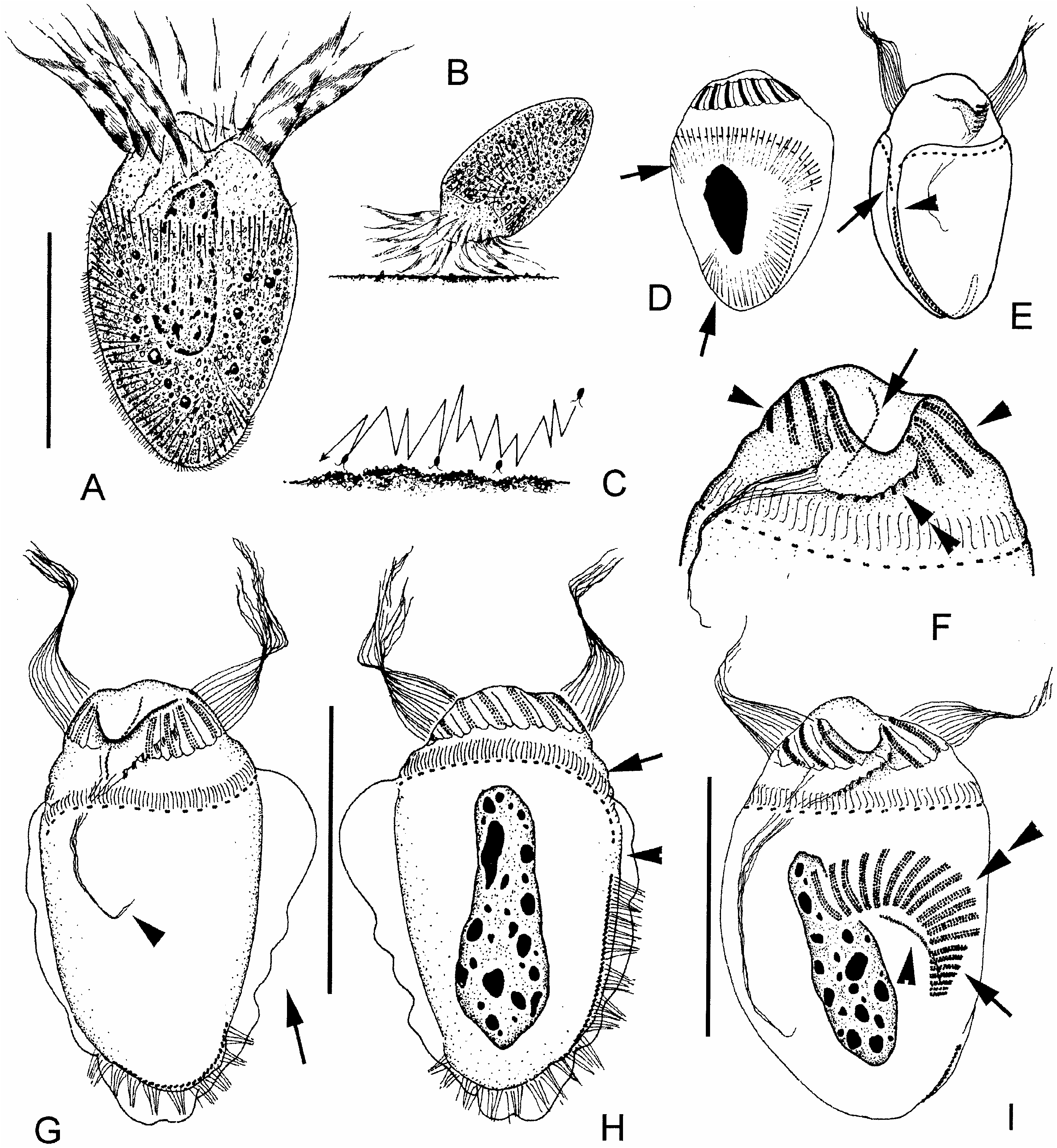
Figure 8. Strombidium apolatum nov. spec. from life (A–C) and after protargol impregnation (D–I). (A) Ventral view of a typical specimen; (B) to show a cell attached to the bottom; (C) pattern of movement; (D) dorsal view, to show the arrangement of extrusomes (arrows); (E) ventral–lateral view, to show the girdle and ventral kinety, which almost meet at equatorial area (arrow and arrowhead, respectively); (F) anterior portion of ventral view, to show the buccal structure: arrow marks the paroral membrane, double-arrowheads indicate the buccal membranelles, arrowheads point to the collar membranelles; note the highly developed cytopharyngeal fibres extending backwards; (G) ventral view, arrow marks the subpellicular platelet layer; (H) dorsal view, arrow marks the equatorial girdle, arrowhead points to the gap between girdle and ventral kinety; (I) an individual in early divisional stage, double-arrowheads mark the newly formed collar membranelles, while arrow indicates the buccal ones; note the paroral membrane (arrowhead). Scale bar: 30 Mm.
Notes
Files
figure.png
Files
(351.7 kB)
| Name | Size | Download all |
|---|---|---|
|
md5:e54f85fc2e7ab63ddbe648df20bf0085
|
351.7 kB | Preview Download |
Linked records
Additional details
Related works
- Is cited by
- Taxonomic treatment: http://treatment.plazi.org/id/294D87A5D362870FFE20DE838C4AFEEF (URL)
- Is part of
- Journal article: 10.1080/00222930400001509 (DOI)
- Journal article: urn:lsid:plazi.org:pub:D574FFDDD3768718FF9CDA548E4CFFEF (LSID)
- Journal article: http://publication.plazi.org/id/D574FFDDD3768718FF9CDA548E4CFFEF (URL)
- Journal article: https://zenodo.org/record/4657793 (URL)