Published April 30, 2005
| Version v1
Figure
Open
Figure 6 in New contributions to the marine benthic ciliates from the Antarctic area, including description of seven new species (Protozoa, Ciliophora)
Creators
Description
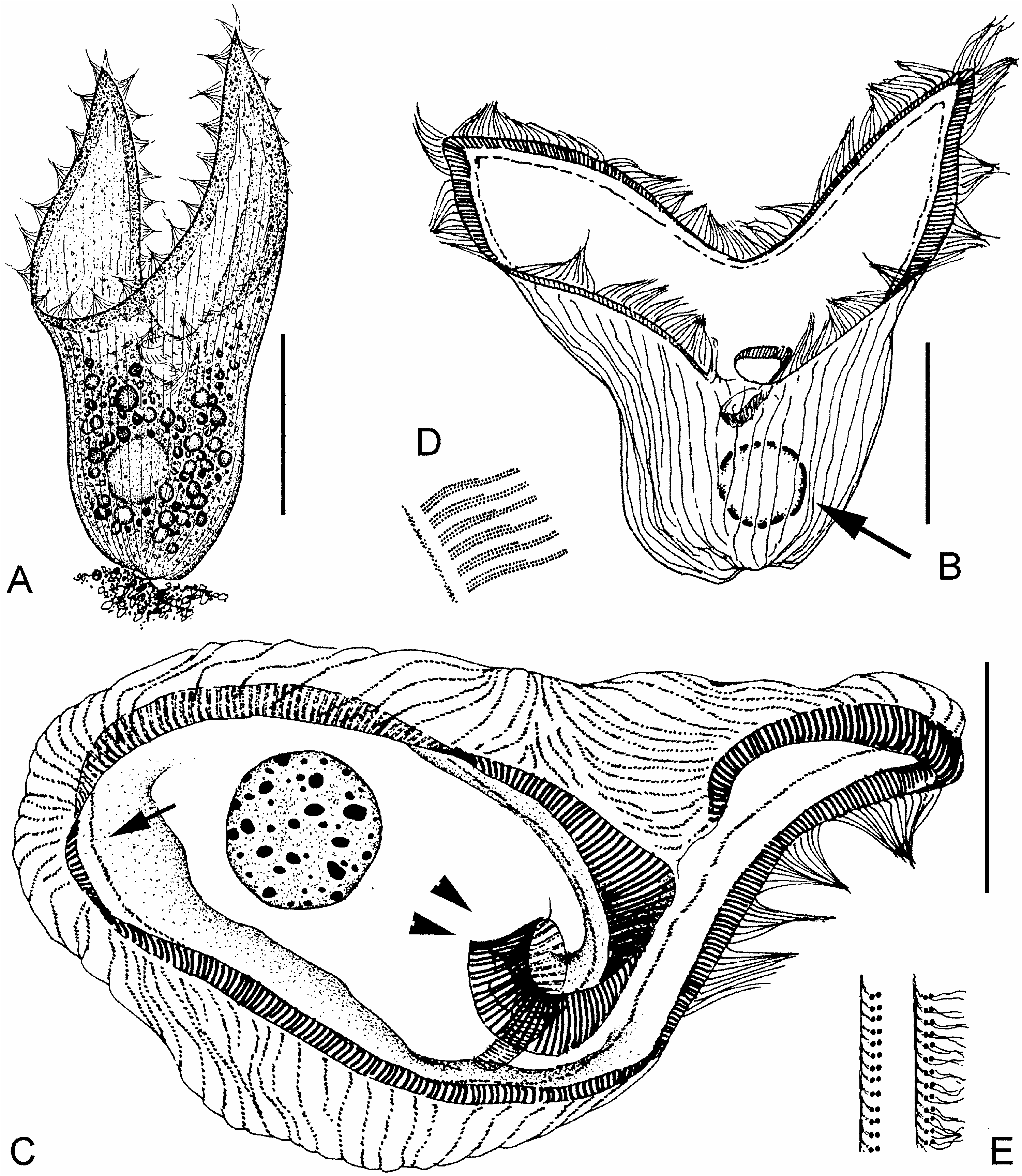
Figure 6. Folliculina? sp. from life (A) and after protargol impregnation (B–E). (A) A typical specimen, note that it is attached to substratum; (B) lateral view, arrow marks the macronucleus; (C) apical view, arrow marks the paroral membrane, arrowheads indicate proximal end of adoral-zone membranelles, which extends deeply into the buccal cavity; (D) details of membranelles and paroral membrane; (E) somatic kineties, to show the fibres associated with dikinetids. Scale bars: 100 Mm (A); 50 Mm (B); 20 Mm (C).
Notes
Files
figure.png
Files
(383.5 kB)
| Name | Size | Download all |
|---|---|---|
|
md5:95c3b47eb72315b64c848aa00b128281
|
383.5 kB | Preview Download |
Linked records
Additional details
Related works
- Is cited by
- Taxonomic treatment: http://treatment.plazi.org/id/294D87A5D3678709FDB9DBE58E9EFB9D (URL)
- Is part of
- Journal article: 10.1080/00222930400001509 (DOI)
- Journal article: urn:lsid:plazi.org:pub:D574FFDDD3768718FF9CDA548E4CFFEF (LSID)
- Journal article: http://publication.plazi.org/id/D574FFDDD3768718FF9CDA548E4CFFEF (URL)
- Journal article: https://zenodo.org/record/4657793 (URL)