Published July 24, 2020
| Version v1
Dataset
Open
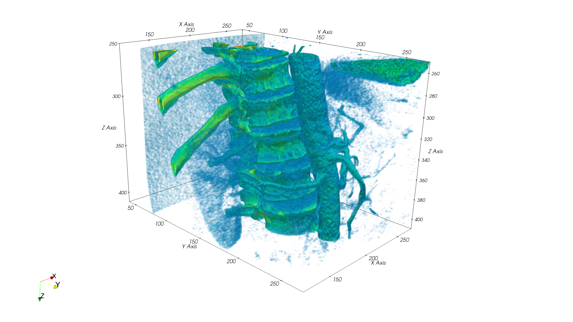
CT image of the upper lumbar and lower thoracic spine
Description
Extract of a computer tomographic imaging dataset of the thorax, showing the upper lumbar and lower thoracic spine, stored as VTK image.
The dataset is used to develop the SODALITE virtual clinical trial use-case. It contains a part of the lumbar spine. Starting caudal at L2 which is only partially contained and ending cranial with T9 which also only partially contained.
The datasets content is illustrated by the attached png image which shows a volume rendering of the dataset.
Format          : VTKFile, type="ImageData", version="1.0"
Header          : ASCII
Byte Order      : LittleEndian
Grid Type       : Rectilinear
Extend  [x,y,z] : 201     , 301     , 161
Spacing [x,y,z] : 0.78125 , 0.78125 , 1.0
Data-Type       : Float64
Min Value       : 0
Max Value       : 2210.5396825
Files
Thorax-RBK_B10f_FoV400_Subset_150-350x50-350x250-410.png
Files
(79.2 MB)
| Name | Size | Download all |
|---|---|---|
|
md5:e1e709270a056035eefed4cf7dc2bfd3
|
1.2 MB | Preview Download |
|
md5:50796da3f11afc7f5364af52e384ac86
|
77.9 MB | Download |