Published December 31, 2015
| Version v1
Figure
Open
FIGURE 3 in Morphology of spermathecae of some pentatomids (Hemiptera: Heteroptera: Pentatomidae) from Turkey
Description
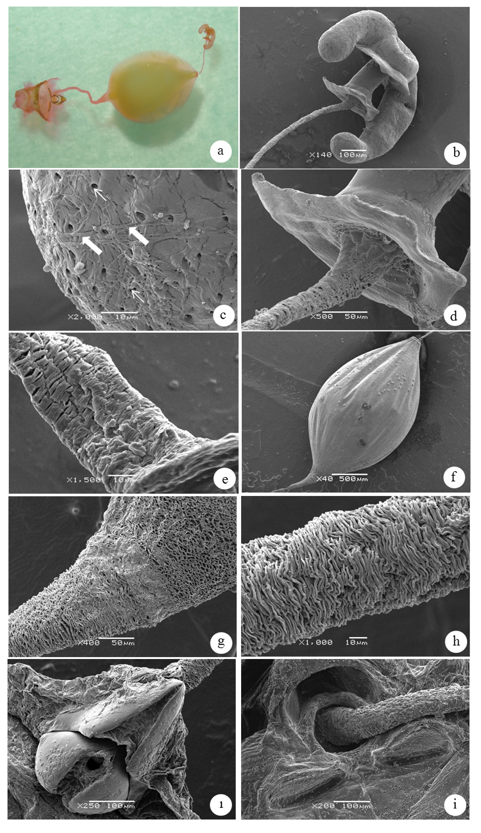
FIGURE 3. The spermatheca of Palomena prasina (Linnaeus) (a). Spermatheca, overview utilizing light microscope. (b). SEM Photo of spermathecal bulb T shaped with two dactyliform like processes. (c). Surface of spermathecal bulb illustrating the arrangement of pores (→) and presence of sperm tails (+). (d–e). Proximal spermathecal flange and distal spermathecal duct. (f). Dilatied surface of spermathecal duct. (g–h). Surface of proximal duct illustrating muscles and spermathecal dilation. (ı). Dorsal surface of genital chamber and (i). Opening of proximal duct.
Notes
Files
figure.png
Files
(4.0 MB)
| Name | Size | Download all |
|---|---|---|
|
md5:65a8bc698030ce9d95fa37a8d38fb981
|
4.0 MB | Preview Download |