Integrated analysis of anatomical and electrophysiological human intracranial data
Creators

- 1. Helen Wills Neuroscience Institute, University of California, Berkeley, Berkeley, CA 94720, USA
- 2. Department of Cognitive Science, University of California, San Diego, La Jolla, CA 92093, USA
- 3. Department of Neurology, University of California, Irvine, Irvine, CA 92697, USA
- 4. Massachusetts General Hospital and Harvard Medical School, Boston, MA 02114, USA
- 5. Radboud University, Donders Institute for Brain, Cognition, and Behaviour, 6500 HB Nijmegen, The Netherlands
Description
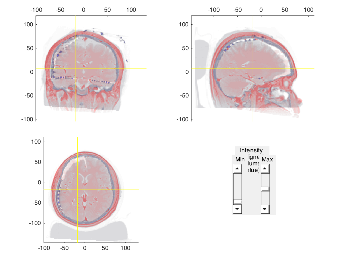
The exquisite spatiotemporal precision of human intracranial EEG recordings (iEEG) permits characterizing neural processing with a level of detail that is inaccessible to scalp-EEG, MEG, or fMRI. However, the same qualities that make iEEG an exceptionally powerful tool also present unique challenges. Until now, the fusion of anatomical data (MRI and CT images) with the electrophysiological data and its subsequent analysis has relied on technologically and conceptually challenging combinations of software. Here, we describe a comprehensive protocol that addresses the complexities associated with human iEEG, providing complete transparency and flexibility in the evolution of raw data into illustrative representations. The protocol is directly integrated with an open source toolbox for electrophysiological data analysis (FieldTrip). This allows iEEG researchers to build on a continuously growing body of scriptable and reproducible analysis methods that, over the past decade, have been developed and employed by a large research community. We demonstrate the protocol for an example complex iEEG data set to provide an intuitive and rapid approach to dealing with both neuroanatomical information and large electrophysiological data sets. We explain how the protocol can be largely automated and readily adjusted to iEEG data sets with other characteristics. The protocol can be implemented by a graduate student or post-doctoral fellow with minimal MATLAB experience and takes approximately an hour, excluding the automated cortical surface extraction.
This collection contains the data described in the protocol and that can be used to replicate all results.
Notes
Files
freesurfer.zip
Files
(572.1 MB)
| Name | Size | Download all |
|---|---|---|
|
md5:65c20ce96ac4cd2c2418e04af7d2a1b5
|
214.6 MB | Preview Download |
|
md5:316dbf06aee9ca37c230eba7a65160a6
|
104.3 MB | Download |
|
md5:014bb7b247e2e4917b81e5c957d678b2
|
187.4 kB | Preview Download |
|
md5:af11c3f32c342327560734172d3700a4
|
197.3 MB | Download |
|
md5:fc9f1368ccba08160f4fb391547b4baa
|
47.4 kB | Download |
|
md5:f0e5967bd8538417e3dd64178aa3a20c
|
53.4 kB | Download |
|
md5:e71adf1781e27833b1c41a38ba35f85a
|
116.7 kB | Download |
|
md5:177186f5b0aeb6d55b2ec2e0c37cc44b
|
54.0 kB | Download |
|
md5:f28965044f8ac8f956a7bddcccd0736d
|
16.1 kB | Download |
|
md5:0ea85e04fc8af7df9e7857f20dce06b8
|
5.3 MB | Download |
|
md5:2f244097d6c726ad6179b3046e5e508a
|
2.8 MB | Preview Download |
|
md5:0dcb7e3c3fa3272de936bae081bca1a4
|
765 Bytes | Download |
|
md5:284f470f1efaefa13c03f2f1092dbb58
|
1.2 MB | Download |
|
md5:7bc567d7c34566cffee38c5749f2b99e
|
23.1 MB | Download |
|
md5:d927574b2fcd8fea7b8913e05a2a12a6
|
23.1 MB | Download |
Additional details
Related works
- Is identical to
- 11633/di.dccn.DSC_3015000.00_734 (Handle)
- Is supplement to
- 10.1101/230912 (DOI)
- 10.1038/s41596-018-0009-6 (DOI)