
Published December 15, 2014
| Version v1
Figure
Open
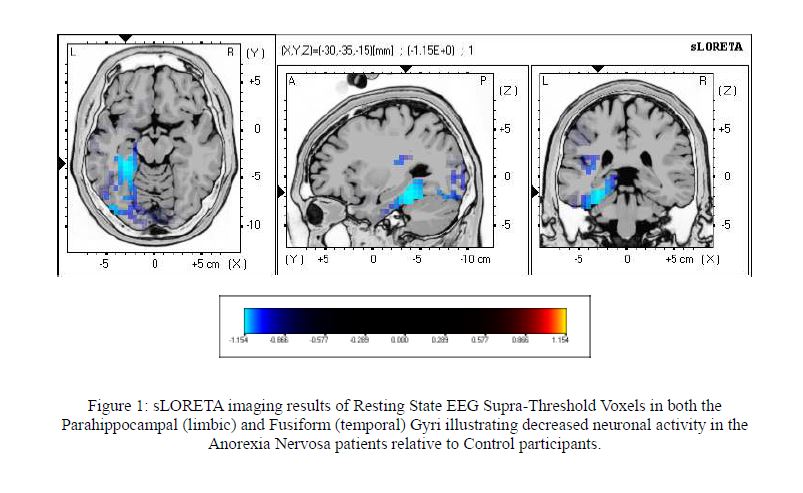
BRAIN Journal-Electrophysiological Neuroimaging using sLORETA Comparing 12 Anorexia Nervosa Patients to 12 Controls-Figure 1: sLORETA imaging results of Resting State EEG Supra-Threshold Voxels in both the Parahippocampal (limbic) and Fusiform (temporal) Gyri illustrating decreased neuronal activity in the Anorexia Nervosa patients relative to Control participants
Creators
- 1. Department of Molecular Pharmacology and Experimental Therapeutics, Division of Clinical Pharmacology, Gonda 19, Mayo Clinic, 200 First Street SW, Rochester, Minnesota 55905, USA
- 2. Neurophysiology Unit, Department of Psychiatry, Medical University of Lublin, ul. Gluska 1 (SPSK Nr 1), Lublin 20-439, Poland
- 3. Department of Electrical Engineering and Measurement Systems, University of Life Sciences in Lublin, 13 Akademicka Street, Lublin 20-950, Poland
Description
The findings of the sLORETA analysis indicated that, the difference is statistically
significant (p=0.03) using a one-tailed t-test: Anorexia > Controls. The brains of the patients with
Anorexia Nervosa illustrated decreased neuronal activity in the Left Fusiform Gyrus and the Left
Parahippocampal Gyrus (p=0.03) in the resting-state brains when Anorexia Patients were sitting for
3min, as compared to the Controls sitting for 3minutes.
Notes
Files
Figure 1. sLORETA imaging results of Resting State EEG Supra.JPG
Files
(80.4 kB)
| Name | Size | Download all |
|---|---|---|
|
md5:e8d7171373b9230f435c841ca615f31f
|
80.4 kB | Preview Download |