Published November 30, 2017
| Version v1
Figure
Open
Figure 11 from: Korshunova T, Martynov A, Bakken T, Evertsen J, Fletcher K, Mudianta WI, Saito H, Lundin K, Schrödl M, Picton B (2017) Polyphyly of the traditional family Flabellinidae affects a major group of Nudibranchia: aeolidacean taxonomic reassessment with descriptions of several new families, genera, and species (Mollusca, Gastropoda). ZooKeys 717: 1-139. https://doi.org/10.3897/zookeys.717.21885
Creators
- 1. Koltzov Institute of Developmental Biology, Moscow, Russia
- 2. Zoological Museum, Moscow State University, Moscow, Russia
- 3. NTNU University Museum, Norwegian University of Science and Technology, Trondheim, Norway
- 4. University Museum, Norwegian University of Science and Technology, Trondheim, Norway
- 5. Unaffiliated, Port Orchard, United States of America
- 6. Universitas Pendidikan Ganesha, Bali , Indonesia
- 7. National Museum of Nature and Science, Tsukuba, Japan
- 8. Gothenburg Natural History Museum, Gothenburg, Sweden|Gothenburg Global Biodiversity Centre, Gothenburg, Sweden
- 9. Biozentrum Ludwig Maximilians University and GeoBio-Center LMU Munich, Munich, Germany|Zoologische Staatssammlung München, Munich, Germany
- 10. Queen's University, Belfast, United Kingdom|National Museums Northern Ireland, Holywood, United Kingdom
Description
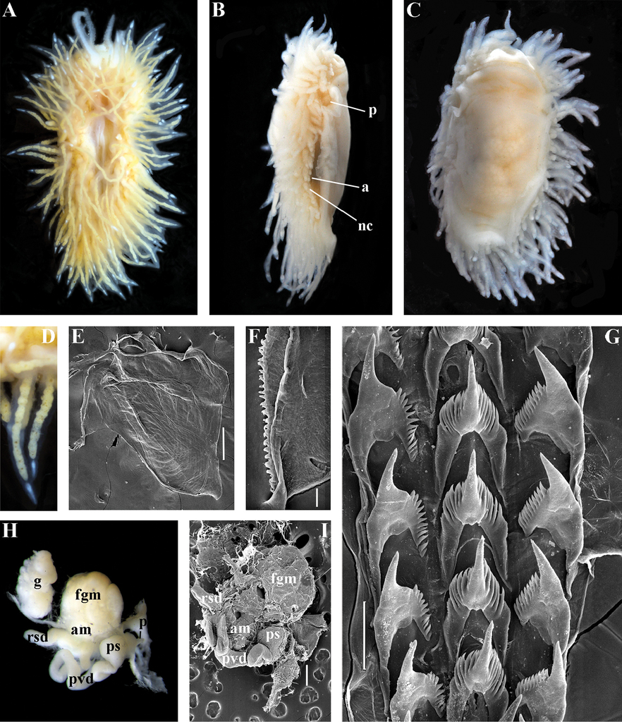
Figure 11 - Polaria polaris (Volodchenko, 1946), comb. n. ZMMU Op-519. Laptev Sea, living specimen 12 mm in length: A dorsal view B latero-ventral view C ventral view D details of cerata E jaw, SEM F details of masticatory process of jaw, SEM G radular teeth, posterior part, SEM H reproductive system, light microscopy I reproductive system, SEM. Abbreviations: a anus am ampulla rsd distal receptaculum seminis fgm female gland mass; g gonad; nc continuous notal edge p penis ps penial sheath pvd prostatic vas deferens Scale bars: E, I = 300 μm; F = 30 μm; G = 100 μm. Photos of living specimens by O.L. Zimina, other photos and SEM images by A.V. Martynov.
Files
big_171529.jpg
Files
(700.5 kB)
| Name | Size | Download all |
|---|---|---|
|
md5:a6bf1185b2cf9aab97dd0de696797815
|
700.5 kB | Preview Download |
Additional details
Related works
- Is part of
- 10.3897/zookeys.717.21885 (DOI)