Published January 31, 2023
| Version v1
Figure
Open
Fig. 6 in Morphology of larval and postlarval stages of Priapulopsis bicaudatus (Danielssen, 1869) (Priapulida) from the north atlantic ocean
Creators
Description
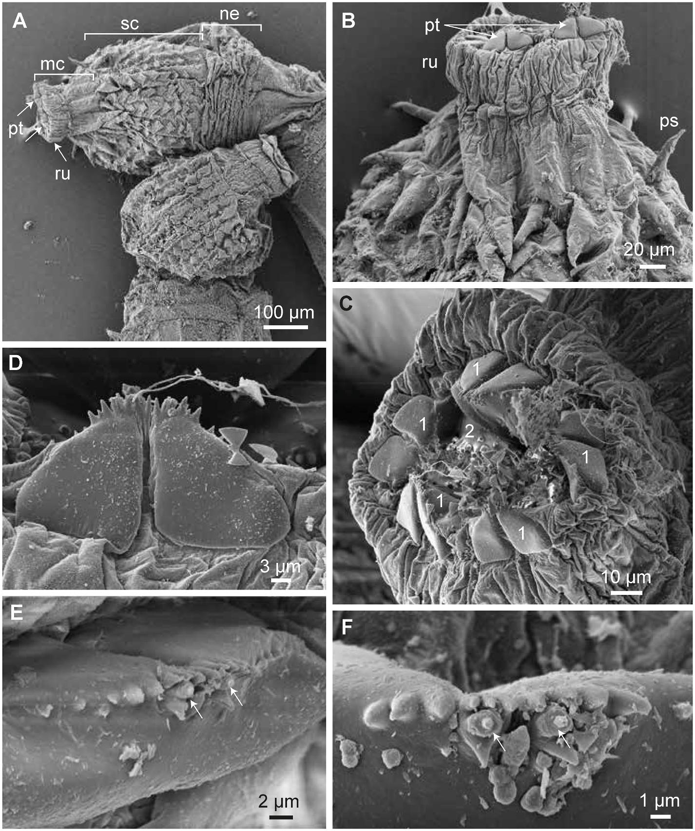
Fig. 6. Pharyngeal teeth in the larva. A. Frontal introvert with widely extended mouth cone (mc), showing teeth of the first ring (pt). B. Teeth (pt) from "outer" side, showing deep median groove. C. Almost frontal view on the first ring of teeth (labelled "1"), part of one tooth of the second ring ("2") is visible. D. Magnification of one tooth from the outer side. E, F. Magnification of the frontal margin with spines and tooth receptors (arrows). Note undivided inner side. Further abbreviations: ne = neck, ps = primary scalid, ru = ruff, sc = scalids. All images SEM, all from V13500.
Notes
Files
figure.png
Files
(2.6 MB)
| Name | Size | Download all |
|---|---|---|
|
md5:de3cd7c126f8aab94ea8dc42985e6ba7
|
2.6 MB | Preview Download |
Linked records
Additional details
Related works
- Is part of
- Journal article: 10.1016/j.jcz.2022.11.006 (DOI)
- Journal article: urn:lsid:plazi.org:pub:FFCBFFC6FFD6C903BE6BFF843115FFC9 (LSID)
- Journal article: https://zenodo.org/record/10375775 (URL)