Evaluation of PD-L1 expression in vulvar cancer
Creators
- 1. (1) Pathology Department, Salah Azaiez Institute, Tunis, Tunisia (2) Research Laboratory LR21SP01, Salah Azaiez Institute, Tunis, Tunisia
- 2. (3) Human Genetics Laboratory LR99ES10, University of medecine of Tunis, University of Tunis El Manar, Tunis, Tunisia
- 3. (1) Pathology Department, Salah Azaiez Institute, Tunis, Tunisia
- 4. (4) Surgical Oncology Department, Salah Azaiez Institute, Tunis, Tunisia
- 5. (2) Research Laboratory LR21SP01, Salah Azaiez Institute, Tunis, Tunisia
Description
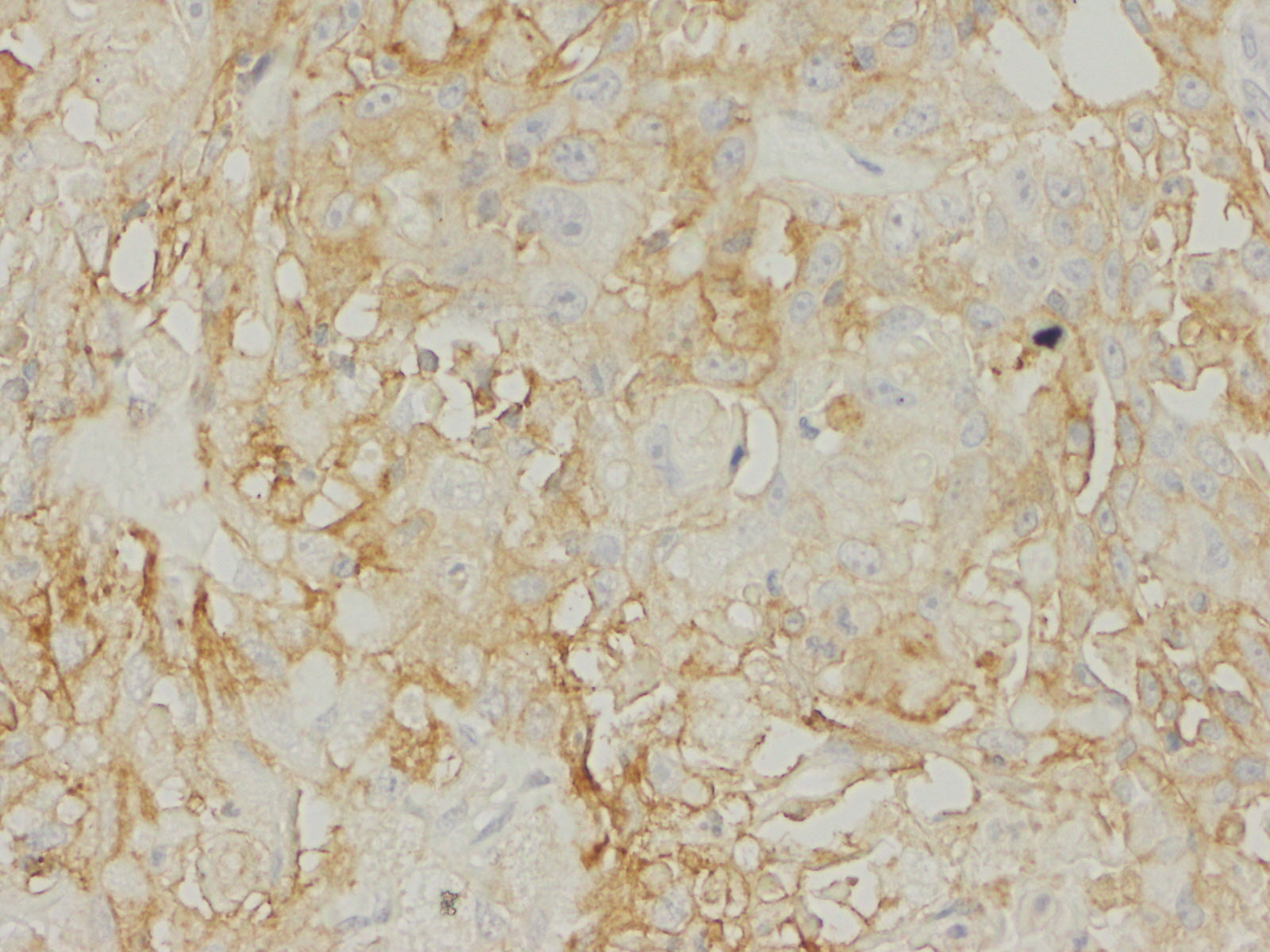
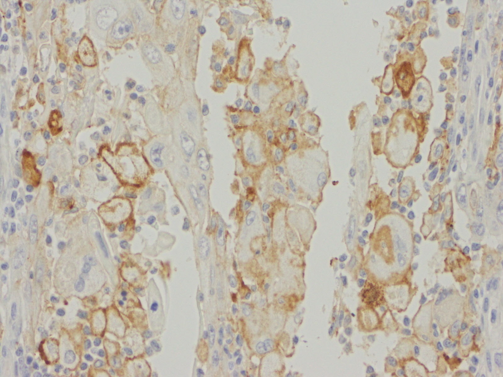
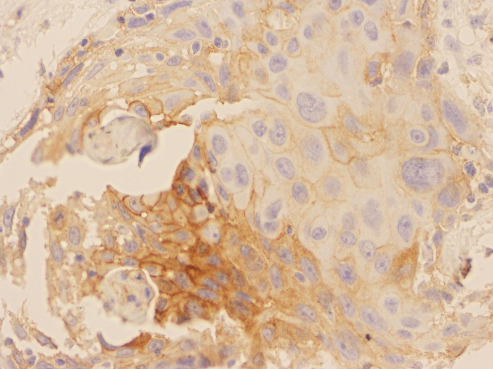
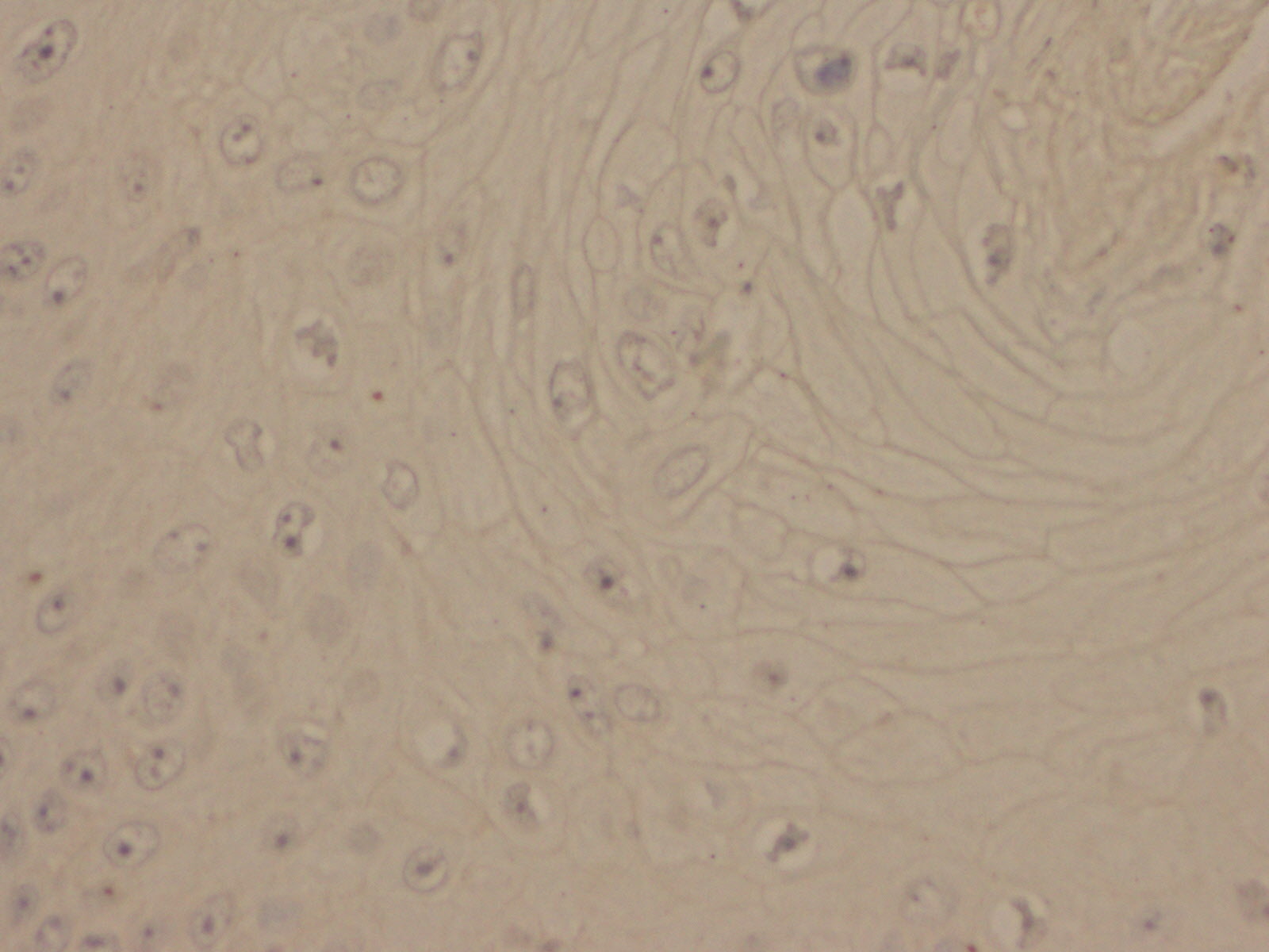
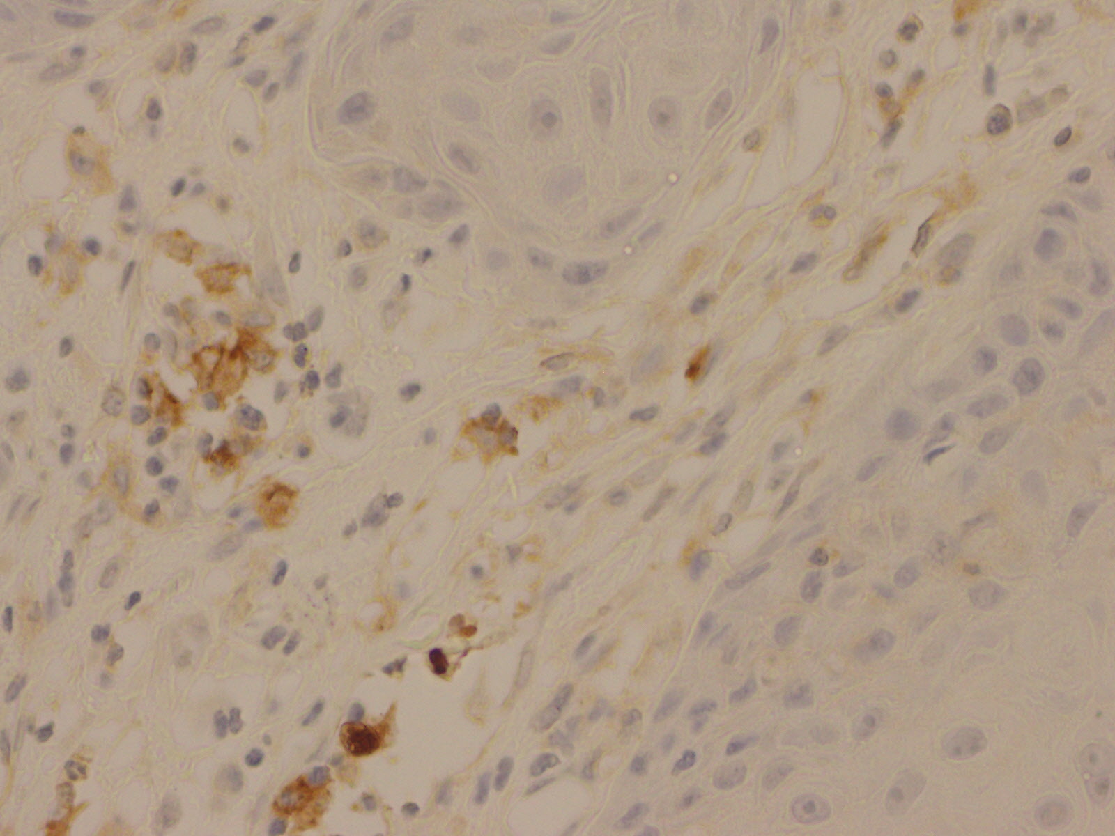
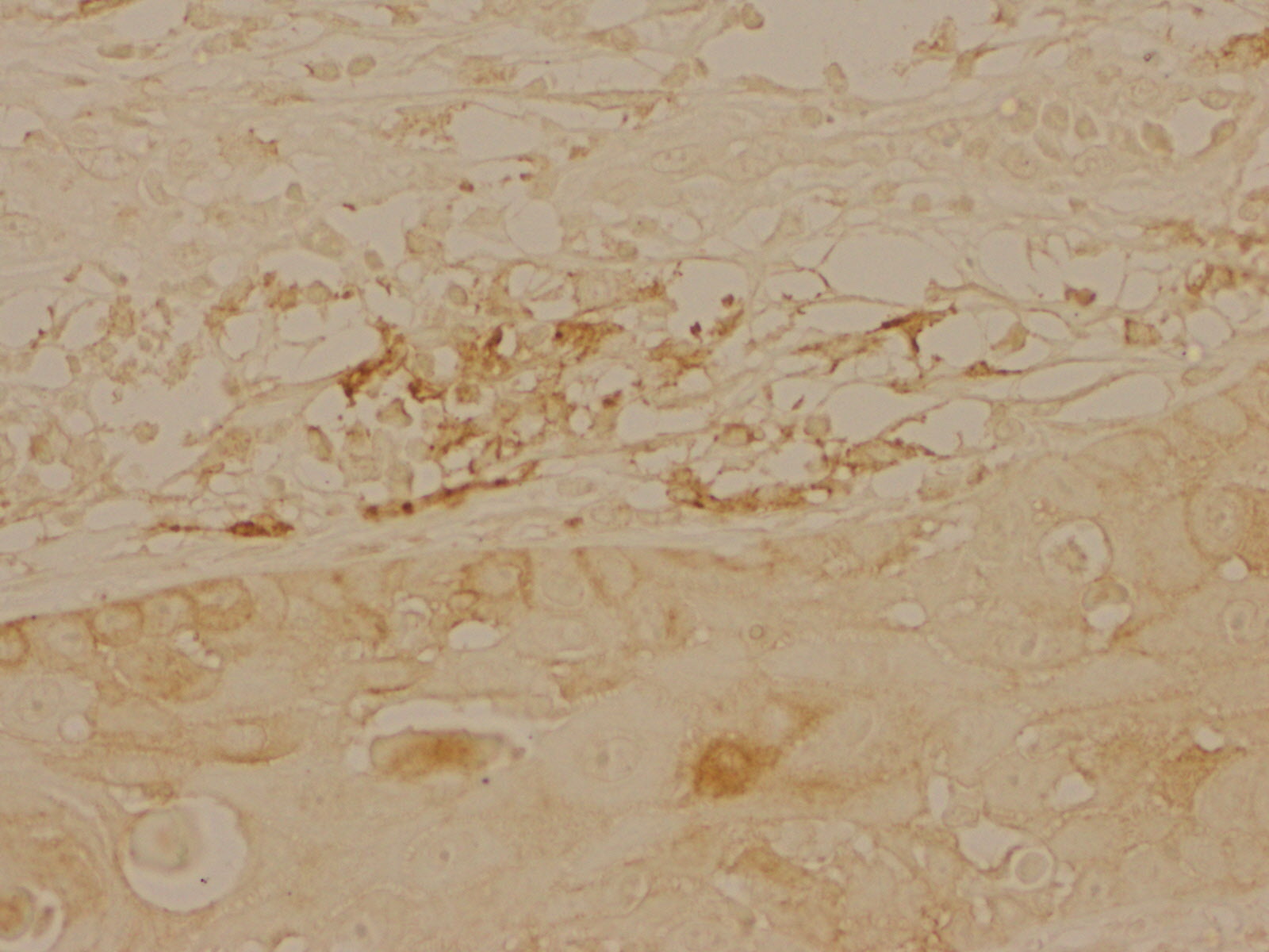
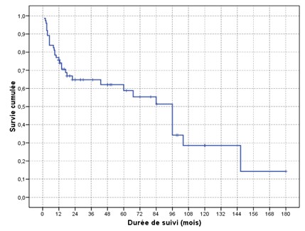
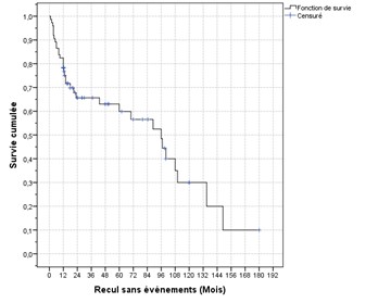
background & Aims: Vulvar cancer accounts for about 4% of all female genital malignancies. Squamous cell carcinoma is the most common histological type.PD-L1 is a membranous protein of nucleated cells. By binding to its PD-1 receptor, PD-L1 inactivates T lymphocytes. It plays a key role in the treatment of some cancers. Our aims were to study the expression profile of PD-L1 in vulvar cancer by immunohistochemistry and to correlate its expression with survival rates. Methods: A retrospective study was conducted in the Pathology Department of Saleh Azaiez Institute, involving 55 patients followed for vulvar cancer over a period of 13 years from January 2008 to December 2021. Clinicopathologic data were collected from medical records and pathology reports. Immunohistochemical analysis was performed using an automaton (Leica Biosystems™). Results: PD-L1 expression in vulvar squamous cell carcinoma was observed in 44% of cases. This expression was noted in 11% of cases at the level of lymphocytes whose dissection was negative in all cases with a maximum tumor size of 40 mm. PDL1 was expressed in tumor cells in 33% of cases where there was a positive inguinal dissection in 22% of cases and a maximum tumor size of 90 mm. Conclusions: The immune checkpoint inhibitor PD-1/PD-L1 seems to have an important prognostic effect on the development of some cancers (breast cancer, lymphoma). Indeed, the expression of PD-L1 in tumors is often considered a factor of poor prognosis due to its immunosuppressive activity in tumor tissues. Many studies have attempted to determine the prognostic factors of vulvar cancer in order to optimize therapeutic management. PD-L1 expression in vulvar squamous cell carcinoma has not been studied before, hence the interest of our study which suggested, as in the literature, a prognostic role for this protein.
Files
fig-1.jpeg
Files
(2.2 MB)
| Name | Size | Download all |
|---|---|---|
|
md5:13f8f143134d3c7a8bc29b9d3cd75d5f
|
442.4 kB | Preview Download |
|
md5:5ca7680c85a5410cba368ed3abf2c4e8
|
397.7 kB | Preview Download |
|
md5:8a4aff63a76017dcaad5b53b5584404b
|
358.4 kB | Preview Download |
|
md5:0ba5a315377104c186acb2f98e9eb852
|
221.5 kB | Preview Download |
|
md5:1e28cca7b4a242c8259d1cb277ac812a
|
307.3 kB | Preview Download |
|
md5:01e82291ac448dd13e95a0e8a57eb403
|
309.2 kB | Preview Download |
|
md5:a3bc04670f0d0942473991f443c74c69
|
32.8 kB | Preview Download |
|
md5:434ae0dd5c516002d5b38a70222ef6ae
|
18.4 kB | Preview Download |
|
md5:c99888f44c14c06bed1a18fc34d8e291
|
6.2 kB | Preview Download |
|
md5:b93360e05f47b1f6a287c4cd43347e37
|
16.0 kB | Download |
|
md5:0d31143ba9f00d49c8008dbe7398aae0
|
40.4 kB | Download |
Additional details
Related works
- Is identical to
- Journal article: 10.1234/foobars.56789 (DOI)