Published September 20, 2021
| Version v1
Figure
Open
FIGURE 3 in Spirobranchus bakau sp. nov. from Singapore: yet another species of S. kraussii-complex (Polychaeta: Serpulidae)
Authors/Creators

- 1. Tropical Marine Science Institute, National University of Singapore, Singapore 119227, Singapore. & tmspssvm@nus.edu.sg; https://orcid.org/0000-0002-4115-292X
- 2. Australian Museum Research Institute, Australian Museum, 1 William Street, Sydney NSW 2010, Australia. elena.kupriyanova@austmus.gov.au; https://orcid.org/0000-0003-0336-4718 & Department of Biological Sciences, Macquarie University, Sydney, NSW 2109, Australia.
- 3. Yale-NUS College, National University of Singapore. Singapore 138527, Singapore. & Department of Biological Sciences, National University of Singapore, Singapore 119223, Singapore.
- 4. Tropical Marine Science Institute, National University of Singapore, Singapore 119227, Singapore. & yapwln@nus.edu.sg; https://orcid.org/0000-0003-4796-1696
- 5. Tropical Marine Science Institute, National University of Singapore, Singapore 119227, Singapore. & tmsteolm@nus.edu.sg; https://orcid.org/0000-0002-3309-4715
Description
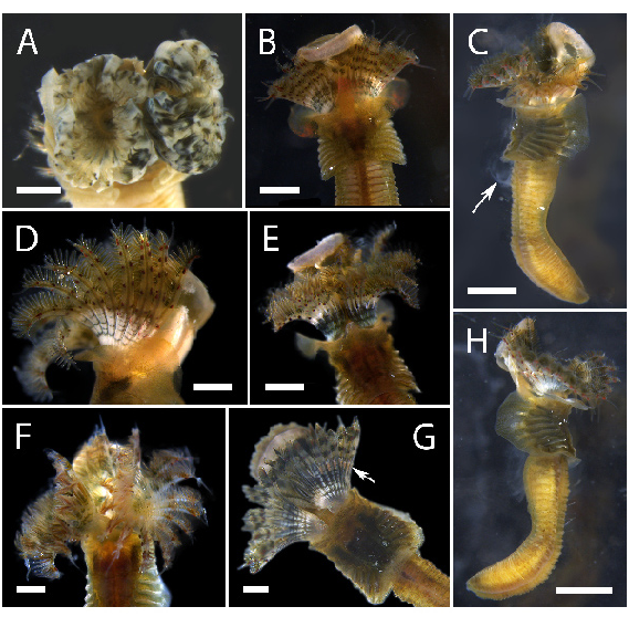
FIGURE 3. Radiolar crown in Spirobranchus bakau sp. nov. specimens. A—Top view of radiolar crown in paratype (ZRC. ANN.0482), showing circular arrangement of radioles; B, D—Radiolar crown with alternating black and cream-coloured bands at the base. The translucent, free ends of the radioles take on the colouration of underlying golden-brown pinnules. Red pigmented spots are mostly arranged in pairs on each radiole; C, H—Lateral views of complete holotype of S. bakau sp. nov. (ZRC.ANN.0480). Image was taken prior to excision of posterior abdominal segments for molecular studies. Evidence of sperm release in C (white arrow), indicating that the specimen is a male; E—Greyish bands alternating with black bands at base of radioles; F—Pinnules near tips are of brownish colouration; G—Distal end of radiolar base in paratype (ZRC.ANN.0481) with peach colouration (white arrow), which extends into inter-radiolar membrane. Scale bars—A–B, D–G: 0.5 mm, C, H: 1 mm.
Notes
Files
figure.png
Files
(624.6 kB)
| Name | Size | Download all |
|---|---|---|
|
md5:019b3d7fafdffa12dcaa58173d8f3d6d
|
624.6 kB | Preview Download |