Published August 1, 2021
| Version v1
Figure
Open
Figure 4 in Postcranial skeletal development of Mugil cephalus (Teleostei: Mugiliformes): morphological and life-history implications for Mugiliformes
Authors/Creators
Description
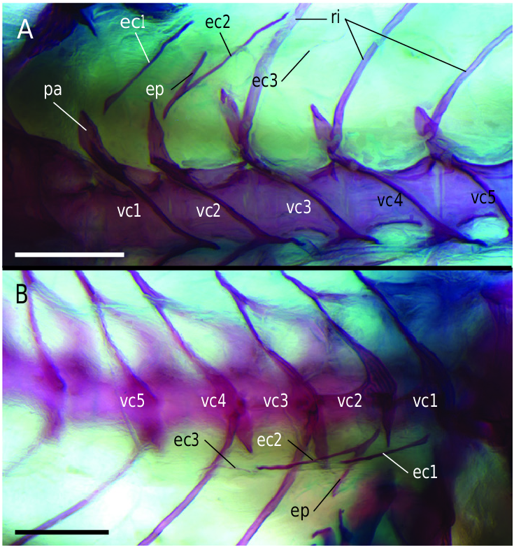
Figure 4. Detailed photographs of the anterior vertebral column of a cleared and stained specimen of Mugil cephalus (DMM IE/16314; L6: 6.5 mm standard length). A, dorsal view. B, dorsolateral view of the right side. Abbreviations: ec, epicentral; ep, epipleural; pa, parapophysis; ri, rib; vc, vertebral centrum. Scale bars: 200 µm.
Notes
Files
figure.png
Files
(1.5 MB)
| Name | Size | Download all |
|---|---|---|
|
md5:d569acc7ed9b59cf635129a1c509a066
|
1.5 MB | Preview Download |
Linked records
Additional details
Related works
- Is part of
- Journal article: 10.1093/zoolinnean/zlaa123 (DOI)
- Journal article: urn:lsid:plazi.org:pub:FFD0FFAC7964F120FFB1C4038D03FF9F (LSID)
- Journal article: https://zenodo.org/record/5301437 (URL)