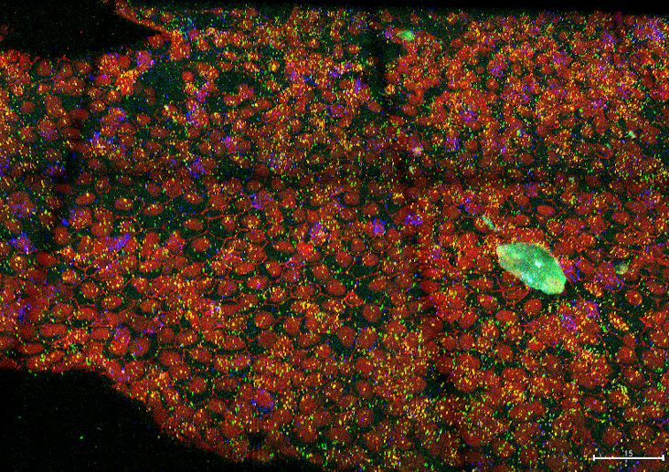
3D confocal images of dorsal views of dissected telencephala from 4 months post-fertilization (mpf) zebrafish, IHC+smFish
Authors/Creators

-
1.
 Centre National de la Recherche Scientifique
-
2.
Institut Pasteur
- 3. CNRS
Description
3D confocal images of dorsal views of dissected telencephala from 4 months post-fertilization (mpf) zebrafish. Dataset of images to complement the paper: FishFeats: streamlined quantification of multimodal labeling at the single-cell level in 3D tissues Bit depth for all the images acquired was 16 bit, a tile scan of multiple Z stacks all with a voxel size of 0.207 µ m by 0.207 µ m by 0.5 µ m. Immunohistochemistry (IHC) for Zo1 (Zonula occludens 1) outlines the apical cell contours, and IHC of Sox2 is used for the identification of neural stem cells and progenitor cells corresponding mostly to the first layer of cells. Expression of pcna, her4 and hey1 (in green, orange and red) were detected using for fluorescent in situ hybridization (FISH)