NFDI4BIOIMAGE Calendar November 2025
Authors/Creators
- 1. Institute of Neurology (Edinger Institute), University Hospital, Goethe University, Frankfurt, Germany
Contributors
Data curator:

-
1.
 Heinrich Heine University Düsseldorf
-
2.
NFDI4BIOIMAGE
Description
Image from the NFDI4BIOIMAGE Calendar November 2025.
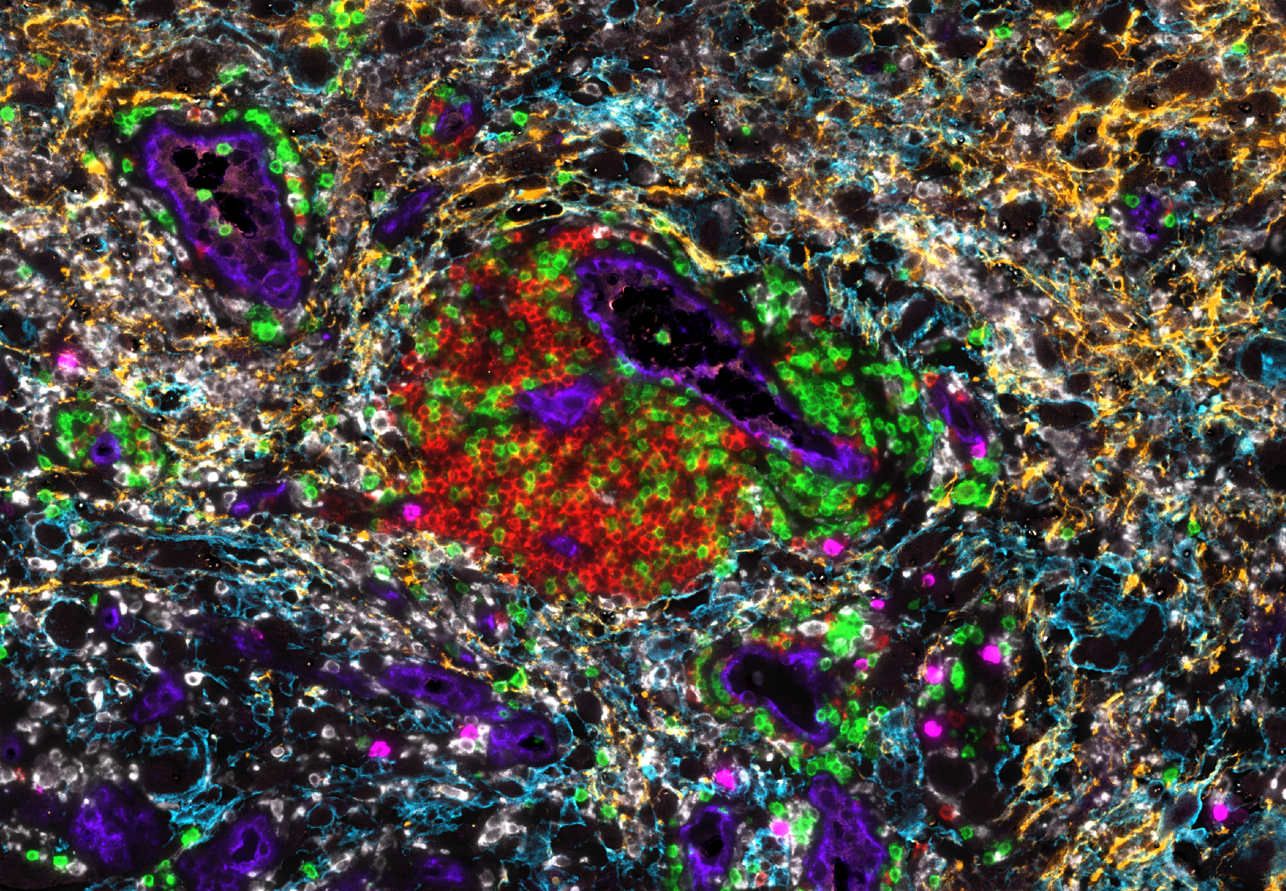
The image shows a perivascular accumulation of B cells, T cells and plasma cells in a human brain tumor. These structures, also known as tertiary lymphoid structures, are sites of lymphocyte clonal expansion and plasma cell formation. The study aims to determine the clinical relevance and immunological function of tertiary lymphoid structures in human primary brain tumors.
Image Metadata (using REMBI template):
|
Study |
|
|
Study type |
Immunomonitoring study in human oncology |
|
Study Component |
|
|
Imaging method |
COMET™ highplex seq-IF staining and scanning system, HORIZON™ Viewer (Lunaphore Technologies, SA) |
|
Biosample |
|
|
Biological entity |
Tertiary lymphoid structure in glioblastoma |
|
Organism |
Homo sapiens |
|
Specimen |
|
|
Location within biosample |
Tumor (glioblastoma) |
|
Preparation method |
FFPE sample, automatic sequential-IF using COMET™ (Lunaphore Technologies, SA) |
|
Signal/contrast mechanism |
HORIZON™ Viewer (Lunaphore Technologies, SA) |
|
Channel 1 - content |
Alexa Fluor Plus 555, red |
|
Channel 1 - biological entity |
CD20 - B-cells |
|
Channel 2 - content |
Alexa Fluor Plus 647, green |
|
Channel 2 - biological entity |
CD3 - T-cells |
|
Channel 3 - content |
Alexa Fluor Plus 555, white |
|
Channel 3 - biological entity |
CD163 - anti-inflammatory macrophages (M2-like) |
|
Channel 4 - content |
Alexa Fluor Plus 647, magenta |
|
Channel 4 - biological entity |
MZB-1 - Marginal zone B and B1 cell-specific protein, MEDA-7 - plasma cells, memory B-cells |
|
Channel 5 - content |
Alexa Fluor Plus 647, orange |
|
Channel 5 - biological entity |
NF- Neurofilament - intermediate filaments around the axons |
|
Channel 6- content |
Alexa Fluor Plus 555, cyan |
|
Channel 6 - biological entity |
GAP43 - Neuromodulin, neuronal growth-associated protein 43 - neurons |
|
Channel 7 - content |
Alexa Fluor Plus 555, blue |
|
Channel 7 - biological entity |
vWF - von-Willebrand-Factor - endothelial cells |
|
Image acquisition |
|
|
Instrument attributes |
COMET™ highplex seq-IF staining and scanning system v.1.1.1.0 (Lunaphore Technologies, SA) |
|
Image acquisition parameters |
COMET™ acquisition software |
|
Image data |
|
|
Pixel size |
0.23 µm/pixel |
|
Image size |
Width 11986 pixels - 2.76 mm |
|
Pixel bit depth |
16-bit |
|
Channel information |
Displayed are 7 markers out of the highplex IF-panel; number of channels 43 (including autofluorescence) |
|
Submitted via NFDI4Immuno |
|
Files
image_november.png
Files
(6.0 MB)
| Name | Size | Download all |
|---|---|---|
|
md5:3490436b96486f36a05648f8228da1bf
|
6.0 MB | Preview Download |
Additional details
Related works
- Is published in
- Preprint: 10.1101/2024.07.04.602038 (DOI)