MR-Eye atlas: a large-scale atlas of the eye based on T1-weighted MR imaging
Authors/Creators
- Barranco Hernandez, Jaime (Researcher)1, 2, 3, 4, 5
- Luyken, Adrian Konstantin (Researcher)6
- Stachs, Philipp (Researcher)7
-
Esteban, Oscar
(Researcher)2, 3

-
Aleman-Gomez, Yasser
(Researcher)2, 3
-
Streckenbach, Felix
(Researcher)8
-
Stachs, Oliver
(Researcher)6, 9
-
Langner, Sonke
(Researcher)6
-
Franceschiello, Benedetta
(Researcher)4, 5
-
Bach Cuadra, Meritxell
(Researcher)1, 3, 2
-
1.
 Centre d'Imagerie BioMedicale
-
2.
Centre Hospitalier Universitaire Vaudois
-
3.
University of Lausanne
-
4.
HES-SO Valais-Wallis
- 5. The Sense Innovation and Research Center
-
6.
Universitätsmedizin Rostock
-
7.
Karlsruhe Institute of Technology
- 8. Department of Ophthalmology, Rostock University Medical Center
-
9.
University of Rostock
Description
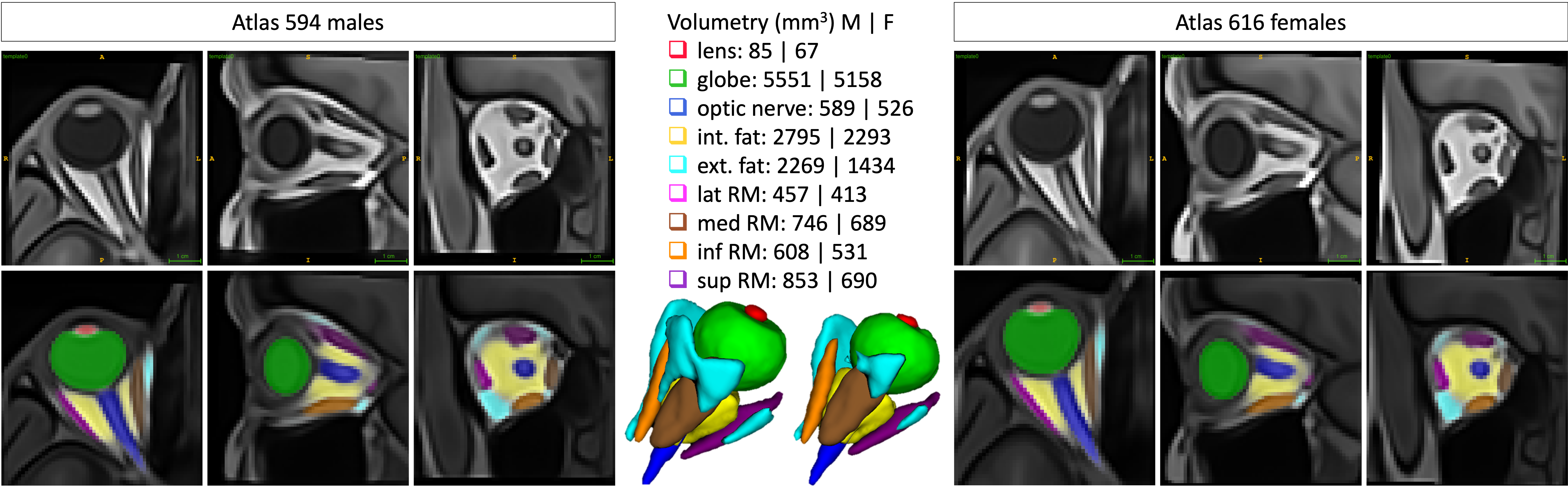
MR-Eye atlas is a novel digital atlas constructed from MR images (T1-weighted MRI acquired at 1.5T) of a large-scale population of healthy volunteers. It gathers male, female, and combined male/female structural atlases constructed from 594 males and 616 females, with corresponding probability maps of the different labels projected onto the average templates. The atlases include 9 regions of interest: lens, globe, optic nerve, intraconal and extraconal fat, and four rectus muscles (lateral, medial, inferior, and superior).
The initial Zenodo release, uploaded on 2024-08-15, included:
- sub_metadata.csv: dataset summary table
- male/0_template.nii.gz and female/0_template.nii.gz: male and female eye atlas images
- male/1_max_prob_map.npy, male/1_max_prob_map.nii.gz, female/1_max_prob_map.npy, and female/1_max_prob_map.nii.gz: maximum probability maps
- male/2_prob_map.npy, male/2_prob_map.nii.gz, female/2_prob_map.npy, and female/2_prob_map.nii.gz: probability maps
Version 2, uploaded on 2025-07-15, added:
- combined/0_template.nii.gz: combined male/female eye atlas image
- combined/1_max_prob_map.npy and combined/1_max_prob_map.nii.gz: maximum probability map for the combined atlas
- combined/2_prob_map.npy and combined/2_prob_map.nii.gz: probability map for the combined atlas
- labels projected onto Colin27 and MNI152 T1w spaces, along with the images and their cropped versions
- additional preview figures showing labels overlaid onto Colin27 and MNI152 images, and a schematic diagram of the processing workflow
- updated README and release notes files
The current update adds:
- combined/3_manual_seg.nii.gz: manual segmentation for the combined eye atlas
- combined/3_manual_seg.itksnap: ITK-SNAP file supporting inspection and editing of the manual segmentation
- updated README and release notes files describing the new manual segmentation files and updated archive structure
Detailed information on the original large-scale cohort, automated segmentation method, unbiased atlas construction, probability maps, and registration to common volumetric coordinate systems is provided in the README file.
MR-Eye atlas is presented and described in the following article:
"A-eye: Automated 3D MRI Segmentation and Morphometric Feature Extraction for Eye and Orbit Atlas Construction", J. Barranco, A. K. Luyken, H. Kebiri, P. Stachs, P. M. Gordaliza, O. Esteban, Y. Aleman, R. Sznitman, F. Streckenbach, O. Stachs, S. Langner, B. Franceschiello, M. Bach Cuadra, https://doi.org/10.1101/2024.08.15.608051
Works using any of the provided resources should cite the above-referenced article.
Copyright (c) - All rights reserved. Medical Image Analysis Laboratory - Department of Radiology, Lausanne University Hospital (CHUV) and University of Lausanne (UNIL), Lausanne, Switzerland & CIBM Center for Biomedical Imaging. 2024.
Notes (English)
Files
A_eye_atlases_male_female.png
Files
(42.6 MB)
| Name | Size | Download all |
|---|---|---|
|
md5:eea40037b70611669211f67aa47dde09
|
1.9 MB | Preview Download |
|
md5:ff306245b18ecf3492e3bd34a5a84d2c
|
5.0 MB | Preview Download |
|
md5:04f948a1d871e7b7e2b653e429ce07bf
|
268.7 kB | Preview Download |
|
md5:88f8908be7d09b0f829c20ae4e67cfe2
|
13.9 kB | Preview Download |
|
md5:034c3085d63dc548e53110b920019449
|
13.6 kB | Preview Download |
|
md5:a812f31e3c0f40c58a0a7a22f22973c8
|
35.4 MB | Preview Download |
|
md5:2ab724713fdaf49e4523c4503bfd068d
|
18.7 kB | Preview Download |
|
md5:8328aaf85594e20ae1f9ecdadbb74c4b
|
718 Bytes | Preview Download |
|
md5:be0e20a96925158e04370b2650177247
|
672 Bytes | Preview Download |


Additional details
Related works
- Is part of
- Preprint: 10.1101/2024.08.15.608051 (DOI)