supplementary figures IRF8 Manuscript
Description
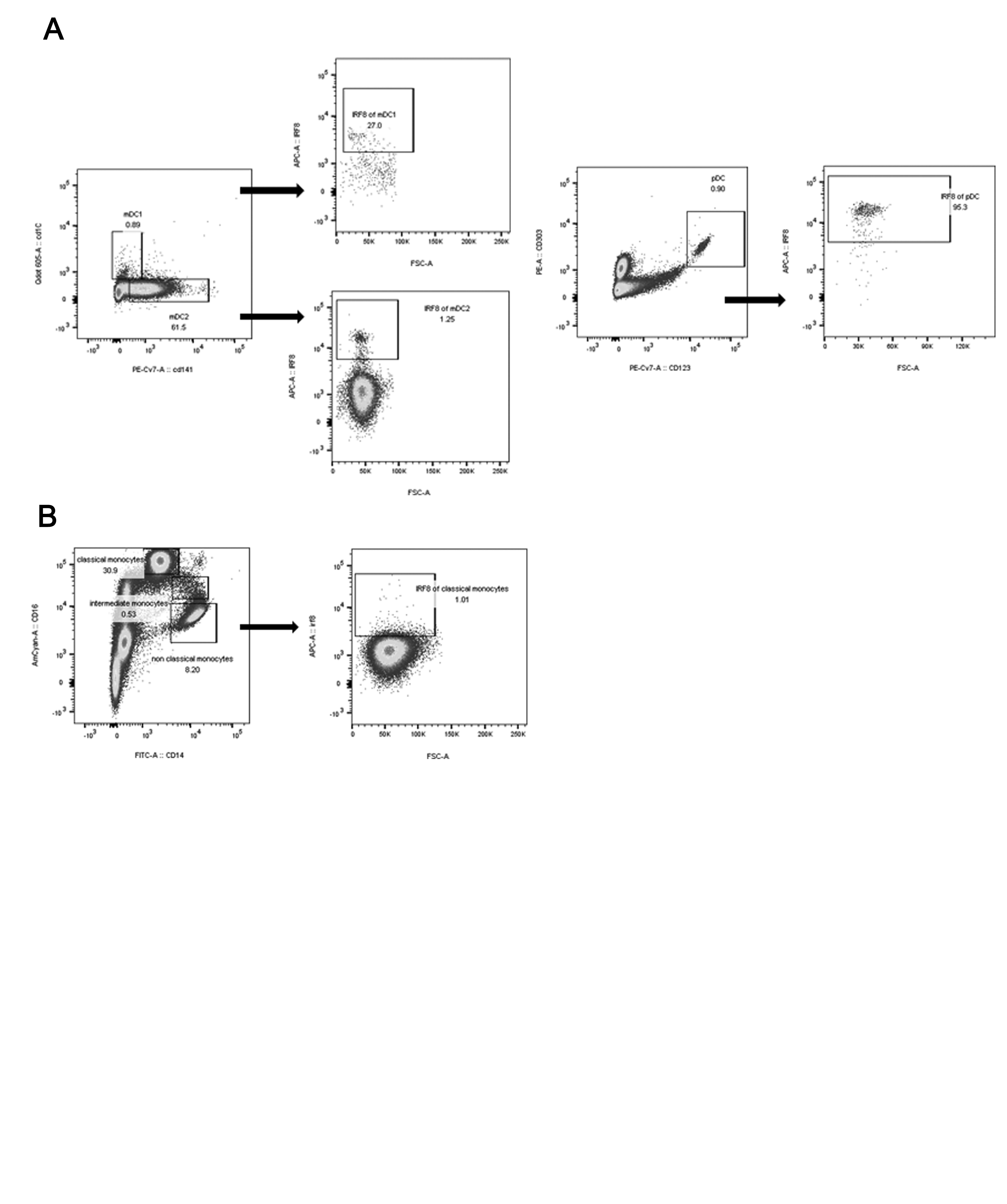
Supplementary Figure 1. Gating strategy for the intracellular staining of IRF8 expression in dendritic cells (A) and monocytes (B).
IRF8, interferon regulatory factor 8; mDC, myeloid dendritic cells; pDC, plasmacytoid dendritic cells.
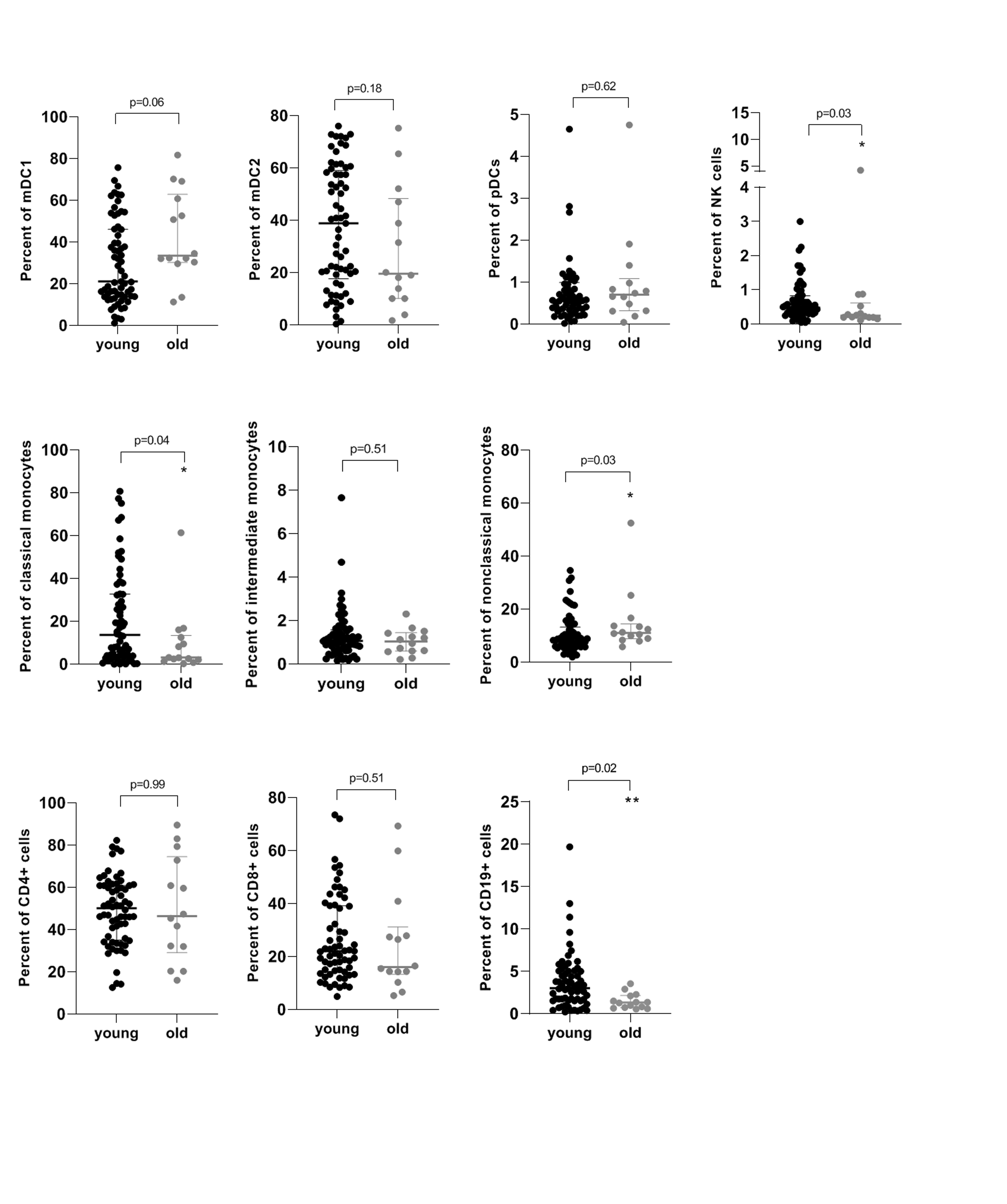
Supplementary Figure 2. Counts of immune cells of innate and adaptive immune system among young and old (>65 years) dialysis patients.
*, p=0.05; **, p=0.01
mDC, myeloid dendritic cells; NK, natural killer; pDC, plasmacytoid dendritic cells.
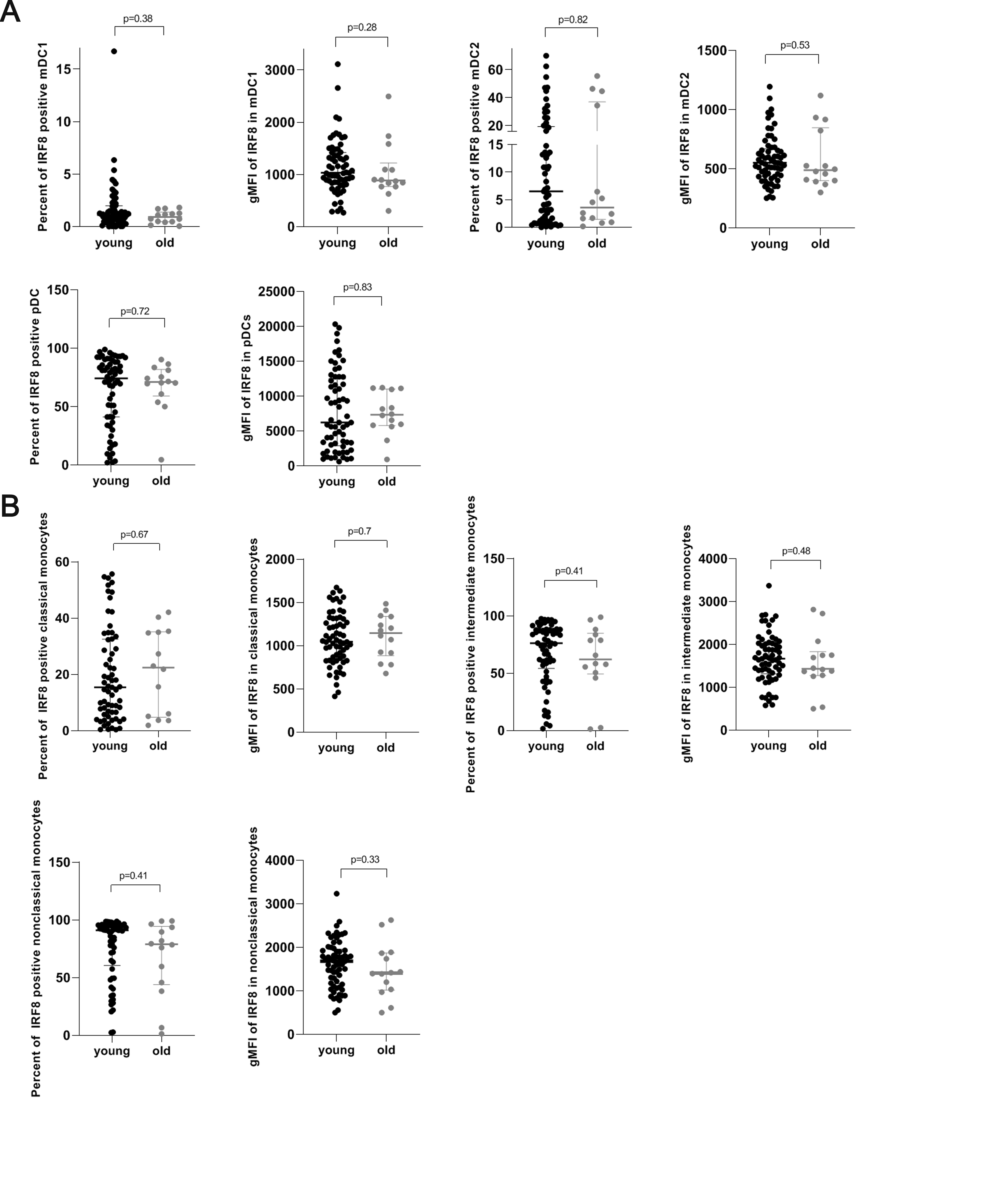
Supplementary Figure 3. Percentages of IRF8 positive dendritic cells (A) and monocytes (B) and IRF8 expression in each dendritic cell and monocyte among young and old (>65 years) dialysis patients.
gMFI, geometric mean; IRF8, interferon regulatory factor 8; mDC, myeloid dendritic cells; pDC, plasmacytoid dendritic cells.