FIGURE 12 in Revision of the Simulium (Simulium) melanopus species-group (Diptera: Simuliidae) in Sabah, Malaysia
Description
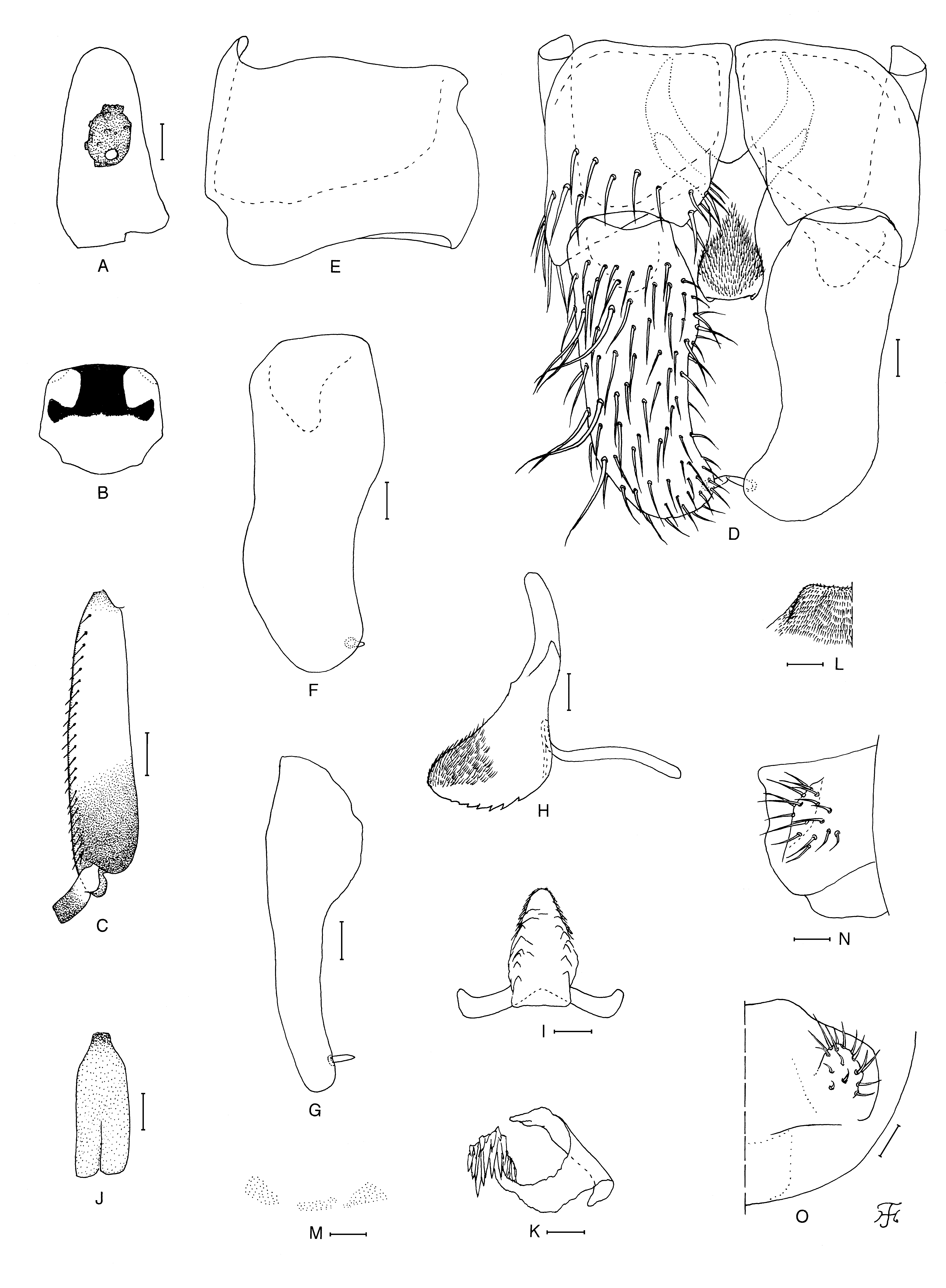
FIGURE 12. Male of Simulium (Simulium) timpohonense sp. nov. (A) Third segment of maxillary palp with sensory vesicle (right side; front view). (B) Scutum showing pruinose pattern (dorsal view). (C) Basitarsus and second tarsomere of hind leg (left side; outer view). (D) Coxites, styles and ventral plate (ventral view). (E) Coxite (right side; ventrolateral view). (F) and (G) Styles (right side; F, ventrolateral view; G, medial view). (H) Ventral plate and median sclerite (lateral view). (I) Ventral plate (caudal view). (J) Median sclerite (caudal view). (K) Paramere (left side; caudal view). (L) Aedeagal membrane (right half; caudal view). (M) Dorsal plate (caudal view). (N) and (O) Tenth abdominal segments and cerci (left side; N, lateral view; O, caudal view). Scale bars. 0.1 mm for C; 0.02 mm for A and D–O.
Notes
Files
figure.png
Files
(592.7 kB)
| Name | Size | Download all |
|---|---|---|
|
md5:b20fb5647b548f07fdc8f50a0abe7501
|
592.7 kB | Preview Download |