Published June 1, 2021
| Version v1
Figure
Open
Figure 6 in Recycling resources: silica of diatom frustules as a source for spicule building in Antarctic siliceous demosponges
Description
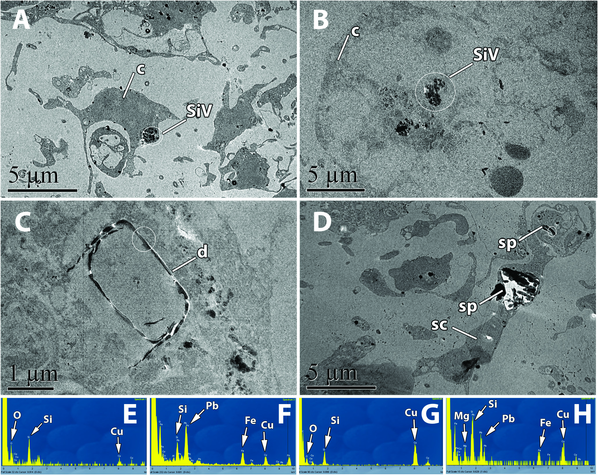
Figure 6. Microanalysis of the content of silica-like vesicles, sponge spicules and diatoms within the sponge tissues. A, sponge cell, probably an amoebocyte (c), of P. areolatus showing accumulation of silica-like granules in the cytoplasm (SiV). B, sclerocyte-like cell (c) of M. tridens showing accumulation of silica-granules in vesicles (SiV). C, diatom (di) engulfed by a sponge cell in M. tridens. D, sclerocyte (sc) of P. areolatus making spicules (sp). E, elemental profile of Figure 6A. F, elemental profile of Figure 6B. G, elemental profile of Figure 6C. H, elemental profile of Figure 6D. Note that the EDX probe measurements were taken on the white circles marked in the images.
Notes
Files
figure.png
Files
(6.4 MB)
| Name | Size | Download all |
|---|---|---|
|
md5:b6c6ec9d075b1796e45babfdd6d50f56
|
6.4 MB | Preview Download |
Linked records
Additional details
Related works
- Is part of
- Journal article: 10.1093/zoolinnean/zlaa058 (DOI)
- Journal article: urn:lsid:plazi.org:pub:FFE4FF83FFEA860FFF130952FFBDFFC3 (LSID)
- Journal article: https://zenodo.org/record/5300095 (URL)